

Biomedical Waste Management Project, Definition, Assignment, PDF
Biomedical waste Management: Any waste generated during the diagnosis, treatment, or immunization of humans or animal is referred to as biomedical waste.Check Biomedical waste Management project here.
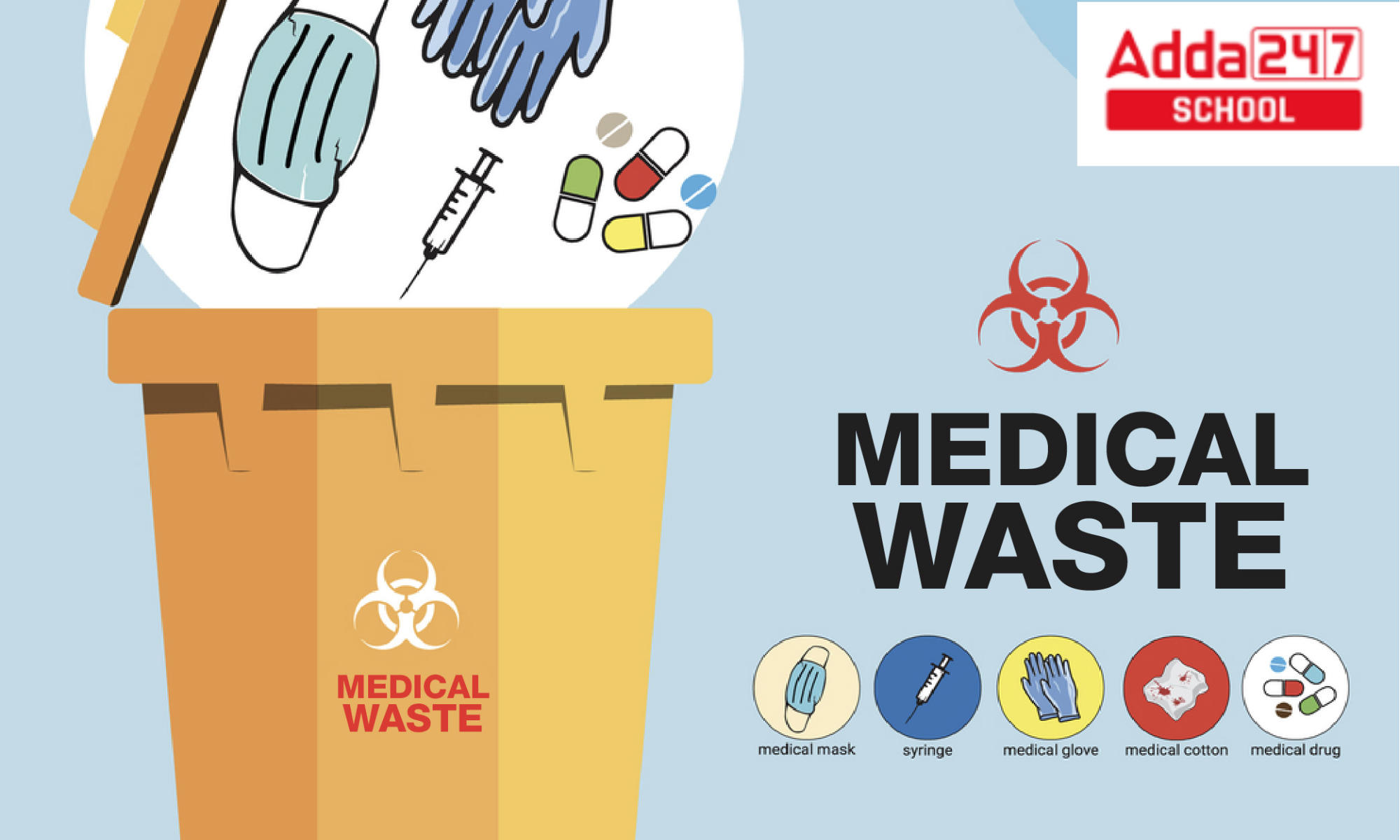
Table of Contents
Biomedical Waste Management
Biomedical waste management involves the proper handling, disposal, and treatment of waste materials generated in healthcare facilities, research laboratories, and other medical settings. It refers to a collection of practises intended to reduce the risks connected with biomedical waste, such as infectious diseases and environmental damage. In this essay, we will look at the significance of biomedical waste management as well as the key principles and practices involved.
Also check, Disaster Management Project for Class 9 &10, PDF Download
Biomedical Waste Management Project Definition
Bio-medical waste management was first governed by the Bio Medical Waste Management and Handling Rule, 1998, and its following changes. The Bio-medical Waste Management and handling regulation 2016 is now in effect. Bio-medical waste is any waste generated during the diagnosis, treatment, or immunization of humans or animals, or during related research activities, as well as the manufacture or testing of biological products in health facilities. The term “bio medical waste” refers to all waste generated by healthcare facilities that, if illegally disposed of, could harm either human health or the environment as a whole. All rubbish that endangers human health or the environment is considered contagious and must be treated as such.
Also Read, Windmill Project for Kids Explanation
Biomedical Waste Definition
Biomedical waste, also known as healthcare waste or medical waste, refers to any waste material that contains biological or infectious agents. This includes discarded items such as used syringes, needles, bandages, laboratory cultures, human tissues, blood, and other bodily fluids. Biomedical waste may also include non-biological materials like chemicals, pharmaceuticals, and radioactive substances used in medical procedures.
Read More, Volcano Project for Kids, Eruption Science Explanation
Importance of Biomedical Waste Management
Proper biomedical waste management is essential for several reasons:
a) Preventing the Spread of Infections: Biomedical waste, especially infectious materials, can harbor pathogens that pose a risk to human health. Effective management practices, such as segregation, disinfection, and proper disposal, minimize the potential for disease transmission among healthcare workers, patients, and the general public.
b) Environmental Protection: Biomedical waste contains hazardous substances that can contaminate soil, water bodies, and the air if not managed properly. By implementing appropriate waste management strategies, the release of toxic chemicals, pathogens, and pharmaceutical residues into the environment can be minimized, safeguarding ecosystems and public health.
c) Compliance with Regulations: Governments and regulatory bodies have established guidelines and regulations for biomedical waste management to ensure public safety and environmental protection. Healthcare facilities are legally obligated to adhere to these regulations to avoid penalties and maintain their reputation.
Also Read, Fluid Mosaic Model of Plasma Membrane, Diagram
Biomedical Waste Management Principle
a) Segregation: The first step in effective waste management is the segregation of different types of biomedical waste. This involves categorizing waste into different color-coded containers based on their characteristics, such as infectious, sharp, chemical, or pharmaceutical waste. Segregation facilitates proper handling, treatment, and disposal of waste materials.
b) Collection and Storage: Biomedical waste should be collected and stored in secure containers that are leak-proof, puncture-resistant, and labeled appropriately. These containers should be placed at designated locations within healthcare facilities to ensure safe and convenient waste disposal.
c) Transportation: Biomedical waste must be transported from healthcare facilities to treatment or disposal facilities in a manner that prevents leakage, spillage, or exposure. Specialized vehicles and trained personnel should be employed for the transportation of biomedical waste, following strict safety protocols.
d) Treatment and Disposal: Biomedical waste requires proper treatment to inactivate pathogens and reduce its potential harm. Common treatment methods include incineration, autoclaving (steam sterilization), chemical disinfection, and microwaving. After treatment, the waste can be disposed of through landfilling, deep burial, or other approved methods.
Read More, Lava Lamp Experiment Ingredients, Aims, Explanation for kids
Best Practices in Biomedical Waste Management
a) Staff Training: Healthcare personnel should receive regular training on the proper handling, segregation, and disposal of biomedical waste. Training should include awareness about potential hazards, infection control measures, and the use of personal protective equipment (PPE).
b) Monitoring and Auditing: Regular monitoring and auditing of biomedical waste management practices ensure compliance with regulations and identify areas for improvement. This includes tracking waste generation, segregation practices, storage conditions, and treatment processes.
c) Public Awareness: Public awareness campaigns can help educate the general population about the proper disposal of biomedical waste generated at home, such as sharps or expired medicines. Clear instructions and convenient collection systems can be provided to encourage responsible waste disposal.
d) Collaboration and Partnerships: Governments, healthcare facilities, waste management authorities, and environmental organizations should collaborate to establish effective biomedical waste management systems.
Biomedical Waste Management Assignment Explanation
Any garbage that contains infectious or possibly contagious elements is considered biomedical waste. These wastes are produced when humans and animals are diagnosed, treated, and immunized. There are both solid and liquid kinds of biomedical waste. Biomedical waste examples include: Waste sharps, including broken glass, scalpels, lancets, syringes, and used needles, bodily parts or recognisable human tissues (as a result of amputation), Veterinary hospital trash and animal tissues, used gloves, dressings, bandages, other medical equipment, contaminated areas’ liquid waste, and waste from the lab. Biomedical wastes must be treated and disposed of differently than ordinary waste.
Types of Biomedical Waste
Biomedical waste is divided into eight categories by the World Health Organization (WHO):
- Infectious Waste
- Sharps objects
- Pathological Waste
- Pharmaceutical Waste
- Genotoxic Waste
- Radioactive Waste
- Chemical Waste
- General/Other Waste
Biomedical Waste Management in Hospital
Biomedical waste in Hospitals include waste from human anatomy, including tissues, organs, and body parts animal waste produced by veterinary hospitals during research, wastes from microbiology and biotechnology, Sharps waste, such as used scalpels, syringes, and hypodermic needles, discarded medications, cytotoxic medications, trash that has been contaminated with blood, such as dressings, bandages, plaster casts, tubes, and catheters, liquid waste from each affected region, and Chemical wastes and incinerator ash.
Utilizing various sorts of containers to collect biological waste from places like operating rooms, labs, wards, kitchens, and hallways is part of the process. It is important to arrange the bins and containers such that 100% collection is obtained.
Biomedical Waste Management Project/ Assignment
Introduction to bio-medical waste (bmw).
All human endeavours result in garbage. We are all aware that this waste could be toxic and needs to be disposed of properly. Polluted water, land, and air, as well as industrial and agricultural waste. Both humans and the environment may be at risk from it.
Similar to this, hospitals and other healthcare institutions produce large amounts of garbage that can spread diseases to anyone who handle it or come into touch with it, including HIV, Hepatitis B and C, and Tetanus.
Definition of BioMedical Waste
Any waste that is produced during the diagnosis, treatment, or immunization of humans or animals, in related research activities, or in the manufacturing or testing of biologicals, as defined by the Biomedical Waste (Management and Handling) Rules, 1998 of India.
Classification of BioMedical Waste
Medical waste is divided into eight categories by the World Health Organization (WHO): general waste, pathological waste, radioactive waste, chemical waste, infectious to possibly contagious waste, sharps waste, pharmaceutical waste, and waste in pressurized containers.
Sources of Biomedical Waste
Hospitals generate waste, which has grown in both quantity and variety over time. In addition to being a risk to patients and the staff who manage them, hospital waste also poses a risk to the environment and public health.
- hospitals, whether public or private, nursing homes, or dispensaries.
- primary care facilities.
- paramedical services, medical schools, and research facilities.
- Animal research facilities and veterinary colleges.
- mortuaries, blood banks, and autopsy facilities.
- Institutes for biotechnology.
- unit of production.
- clinics for doctors and dentists.
- slaughterhouses for animals.
- camps for blood donation.
- vaccine facilities.
- Psychiatric facilities, cosmetic piercing, and acupuncturists.
- funeral arrangements.
- institutions for people with disabilities.
Treatment of Biomedical Waste Management
1. chemical processes.
These procedures make use of disinfectant-acting compounds. Examples of such compounds include ozone, hydrogen peroxide, per-acetic acid, sodium hypochlorite, dissolved chlorine dioxide, and dry inorganic chemicals. The majority of chemical reactions require neutralising agents and a lot of water.
2. Thermal Processes
Heat is used in these procedures to disinfect. Low-heat systems and High-heat systems have been divided into two categories based on the temperature at which they function. Steam, hot water, or electromagnetic radiation are used in low-heat systems (which run between 93 and 177°C) to heat and cleanse the waste.
3. Mechanical Processes
To make trash handling easier or to process waste in conjunction with other treatment stages, these methods are used to alter the physical shape or features of the waste. The key two mechanical operations are
- Compaction: used to lessen the amount of waste
- Shredding: used to prevent the reuse of plastic and paper waste by destroying it. A shredder can only be used with waste that has been disinfected.
4. Irradiation Processes
Wastes should be exposed to ultraviolet or ionising radiation in a sealed space. To make the garbage unidentifiable in these systems, post shredding is necessary.
5. Biological Processes
treating medical waste with biological enzymes. In addition to decontaminating the waste, it is asserted that biological reactions will also cause the elimination of all organic components, leaving only plastics, glass, and other inert materials in the residues.
Health Hazards of Biomedical Waste Management
The WHO reports that the average life expectancy in the world is rising. However, the number of fatal infections is rising. According to a WHO research, infectious diseases claim the lives of more than 50,000 people every day.
Ineffective waste management is one of the factors contributing to the rise in infectious diseases. The majority of viruses, bacteria, and parasites that cause illness are found in blood, body fluids, and bodily secretions, which are components of biomedical waste.
Biomedical Waste Management Assignment Project PDF
Here we give bio medical waste management assignment project pdf for the students. check now
Bio Medical Waste Management
- Health Ministers Of India List From Independence To 2022
- How Many Liters In A Gallon Of Water?- In India, UK, US
- Sustainable Development Project, Definition, Example, PDF For Class 10
- Oscillatory Motion, Meaning, Definition, Example, Diagram
- SSC Full Form In 10th School Education In Hindi And English
- Heterogeneous Mixture- Meaning, Definition & Example For Class 9
- My Best Friend Essay In English 200 Words For Boys/Girls PDF
- Important Question Of Physics Class 12 With Answers
- Parenchyma- Cells, Tissue, Meaning, Function, And Diagram
- What Is Hybridization?- Sp3, Sp2, Examples And Formula
Sharing is caring!
What are the 4 types of biomedical waste?
Infectious, hazardous, radioactive, and normal medical waste are the four main categories.
What are the types of biomedical waste management?
Incineration is one of the several technologies that can be utilised for treatment. Chemical eradication, Thermal Wet Treatment, Radiation from microwaves, Disposal of Land, and Inertization.
What is biomedical waste and its types classify?
There are two categories for biomedical waste: 2. Hazardous garbage, followed by non-hazardous waste Non-hazardous waste: Page 5 About 75% to 90% of the properties of biomedical waste are identical to those of household garbage and are not dangerous in any way.
What is called biomedical waste?
Any waste generated during the diagnosis, treatment, or immunisation of humans or animals used in research operations, the manufacture or testing of biological products, or in health camps is referred to as biomedical waste (BMW).

Leave a comment
Your email address will not be published. Required fields are marked *
Save my name, email, and website in this browser for the next time I comment.
Trending Articles
- NEET PG Admit Card 2024
- CUET Result 2024
- CBSE 10th Compartment Result 2024
- NEET Syllabus 2025
- NEET Counselling 2024
- NEET Cut Off 2024

CBSE Board Exam 2024
- CBSE Previous Year Papers
- CUET Syllabus
- CUET Previous Year paper
- CUET Participating College & Universities
- JEE Main Syllabus 2025
- NEET State wise Cut off
- NEET Rank Predictor
- NEET OMR Sheet
- NEET College Predictor
Recent Posts
- National Symbols of India with Names for Kids in Chart
- Computer Full Form, Check 70 Full Form of Computer in English
- 12 Months Name in English for January to December
- 100 Flowers Names List in English and Hindi, Download PDF
- What are the 11 Fundamental Duties of Indian Constitution?
- Pollution Essay in English [1000 Words]
- Rain Water Harvesting Project, Definition, Methods for Class 10
- Father Of Mathematics- Check Who is the Father of Maths in India
- Largest Delta in India- Know The Longest Delta in the World
- 118 Elements and Their Symbols and Atomic Number and Mass Number PDF
- 100 Vegetables Name in English and Hindi with Pictures
- Who Invented Exams?- Henry Fischel
- River Map of India- Check Indian River Map with Names
- Country Name- All Total 195 Countries in World List
- Important Festivals of India State wise List, Check Top 10 Festival Names
- Mahatma Gandhi Essay 10 Lines in English 300 Words
- Coldest Place in India- Know Lowest Temperature State in India
- Which is the Ugliest Language in India and World?
- How Many Countries are There in Asia?
- 10 Input and Output Devices of Computer Chart
- How Many Districts are There in India 2024
- Lakes in India- Know Important Lakes of India List
- Bank Statement Application to Bank Manager
- Fruits Name in English and Hindi
- 8 Planets Names in English
- Physics Formulas, Check All Basic Important Formula Sheet Class 12
- Keyboard Shortcut Keys of Computer A to Z PDF
- भारत का नक्शा-Bharat ka Naksha | India Map with States
- Acetone Formula, Structure, Name, Uses, Reactions in Chemistry
- Velocity – Definition, Formula, Units, Equation, Examples
IMPORTANT EXAMS
Ncert solutions.
- NCERT Class 12
- NCERT Class 11
- NCERT Class 10
- NCERT Class 9
NCERT Books
School syllabus.
- CBSE Class 12
- CBSE Class 11
- CBSE Class 10
- CBSE Class 9
- JEE Mains 2024
Our Other Websites
- Teachers Adda
- Bankers Adda
- Current Affairs
- Adda Bengali
- Engineers Adda
- Adda Marathi
- Adda School

Get all your queries solved in one single place. We at Adda247 school strive each day to provide you the best material across the online education industry. We consider your struggle as our motivation to work each day.
Download Adda247 App
Follow us on
- Responsible Disclosure Program
- Cancellation & Refunds
- Terms & Conditions
- Privacy Policy
- DOI: 10.7759/cureus.34589
- Corpus ID: 256591284
Biomedical Waste Management and Its Importance: A Systematic Review
- Himani S Bansod , Prasad Deshmukh
- Published in Cureus 1 February 2023
- Medicine, Environmental Science
Figures from this paper


9 Citations
Navigating challenges in biomedical waste management in india: a narrative review, exploring biomedical waste management practices among healthcare professionals: a study from a tertiary care teaching hospital in eastern india, the efficient disposal of biomedical waste is critical to public health: insights from the central pollution control board guidelines in india.
- Highly Influenced
Healthcare Workers’ Knowledge about the Segregation Process of Infectious Medical Waste Management in a Hospital
The medical waste management issues, challenges and solution in indian perspective., simulation clinic waste audit assessment and recommendations at the university of washington school of dentistry., performance evaluation of the sterilization process with bowie & dick test and biological indicator in the quality control of a blood bank in peru, defining green gap in environmental physiotherapy: challenges, advocacies and solutions, combining patient care and environmental protection: a pilot program recycling polyvinyl chloride from automated peritoneal dialysis waste, 36 references, bio medical waste management and its treatment.
- Highly Influential
Bio-medical waste disposal in India: From paper to practice, what has been effected
A study of awareness about biomedical waste management among health care personnel, biomedical waste management in india: a review, biomedical waste management in india: critical appraisal, a study on evaluation of biomedical waste management in a tertiary care hospital in south india, nanoparticles impact in biomedical waste management, health hazards of medical waste and its disposal, medical laboratory waste generation rate, management practices and associated factors in addis ababa, ethiopia, biomedical waste management : an infrastructural survey of hospitals., related papers.
Showing 1 through 3 of 0 Related Papers
- Increase Font Size
21 Biomedical wastes: Definition, sources, classification, collection, segregation, Treatment and disposal
Dr. J. Rajesh Banu
1.Objectives:
- To know what is biomedical waste and its source of generation To gain knowledge about the different types of biomedical waste. To explain the steps of biomedical waste management
- To describe the proper mode of collecting and segregating the biomedical waste To understand the risks of biomedical waste, its method of treatment and disposal
2. Biomedical Waste: Definition:
Bio-medical waste means “any solid and/or liquid waste produced during diagnosis, treatment or vaccination of human beings or animals. Biomedical waste creates hazard due to two principal reasons: infectivity and toxicity. Figure 1 shows some of the biomedical waste
Figure 1.Biomedical waste
3. Sources:
The source of biomedical waste is the place or the location at which biomedical waste has been generated. The source of biomedical waste is classified into two types based on the quantity of waste generated. They include major and minor source. Major source generates more amount of biomedical waste compared to minor source and also there is regular generation of biomedical waste in the major source which includes government hospitals, private hospitals, nursing home and dispensaries. Minor source includes physicians and dental clinics. Figure 2 shows the details of the various source of biomedical waste generation
Figure 2 Sources of biomedical waste
4. Classification:
The classification of the biomedical waste is carried out based on its characteristics, source of generation and the level of hazard to the environment. The biomedical waste is classified into two types:
1. Non hazardous waste
2. Hazardous waste
4.1 Non-hazardous waste:
About 75% to 90 % of biomedical waste characteristics were similar to that of domestic waste and are non-risky in nature. This waste is generated mainly from the organization and maintenance of hospital and health care centers.
4.2 Hazardous waste:
The remaining 10 – 25% of biomedical waste falls under the hazardous waste categories. The hazardous waste contains infectious characteristics of about 15% – 18 % and toxicity characteristics of about 5% – 7%. The various hazardous wastes includes,
Infectious waste: Waste containing pathogens; e.g. excreta; laboratory cultures; isolation wards waste; swabs, materials, or equipments that have been in contact with infected patients.
Pathological waste: Human tissues or fluids e.g. body parts; blood and other body fluids; fetuses.
Pharmaceutical waste: Waste containing pharmaceuticals; e.g. pharmaceuticals that are expired or no longer needed; contaminated pharmaceuticals (bottles, boxes).
Genotoxic waste: Waste containing cytostatic drugs (often used in cancer therapy)/ genotoxic chemicals.
Chemical waste: Waste containing chemical substances e.g. laboratory reagents; film developer; disinfectants and solvents that are expired or no longer needed.
Wastes with high content of heavy metals: Batteries, Broken thermometers, blood pressure gauges, Pressurized containers, gas cylinders, gas cartridges, aerosol cans.
Radioactive waste from radiotherapy: Waste containing radioactive substances e.g. unused liquids from laboratory research; contaminated glassware, packages or absorbent paper; urine and excreta from patients treated or tested with uncapped radionuclide
5. Biomedical Waste management:
Proper management of biomedical waste is highly essential since it induces various risk to the human health and to the surrounding ecosystem that leads to the ecological hazard, professional hazard and public hazard. Steps involved in biomedical waste management was shown in Figure 3
Figure 3. Steps involved in biomedical waste management
5.1 Segregation
To avoid mixing of the biomedical waste with other, a container should be set to the side with colour coding bags at the point of generation. The sorting or separation of waste into different categories is referred as segregation. Segregation will decrease or minimize the risks in addition to rate of managing and disposal. Segregation is the most important and critical step in bio-medical waste management. Only, effective segregation can confirm the effective bio-medical waste management.
5.1.1 How does segregation help?
Segregation plays an effective role in handling and treatment of waste. It reduces the quantity of waste and if done effectively, it can avoid the mixing of biomedical waste with any other type of waste especially municipal waste. Segregation will avoid the reuse of certain biomedical waste like used syringes, needles and other plastics. Some materials like plastics can be recycled after proper disinfection and these can be reused for non-food grade products.
During segregation process, the biomedical waste must be separated under the following categories shown in Table 1. Category no.1 includes the Human anatomical waste in which the human tissues, organs, body parts are considered. Animal waste falls under the Category No. 2. It includes Animal tissues, organs, body parts, carcasses, bleeding parts, fluid, blood and experimental animals used in research, waste generated by veterinary hospitals and colleges, discharges from hospitals, animal houses. Category No. 3 is the Microbiology & Biotechnology waste which contain Wastes from laboratory cultures, stocks or specimen of live microorganisms or attenuated vaccines; human and animal cell cultures used in research; infectious agents from research and industrial laboratories; wastes from production of biologicals, toxins and devices used for transfer of cultures. The Category No. 4 includes waste Sharps in which Needles, syringes, scalpels, blades, glass, etc. that may cause puncture and cuts. This includes both used and unused sharps. Discarded Medicine and Cytotoxic drugs falls under the Category No 5 which consists of wastes comprising of outdated, contaminated and discarded medicines. The soiled waste is included in the Category No. 6 containing items contaminated with body fluids including cotton, dressings, soiled plaster casts, lines, bedding and other materials contaminated with blood.
Table 1. Categories of Waste (Source: Biomedical Waste (Handling and management Rules 1998)
Category No 7 is the solid waste which includes waste generated from disposable items other than the waste sharps such as tubing, catheters, intravenous sets, etc. Liquid waste falls under the category no. 8, it consists of waste generated from the laboratory and washing, cleaning, housekeeping and disinfecting activities. Category No 9 includes incineration ash i.e., ash from incineration of any biomedical waste. Chemical Waste falls under Category No 10 and consists of Chemicals used in production of biologicals, chemicals used in disinfection and as insecticides etc.
5.2 Collection and storage
The collection of biomedical waste involves the installation of different colour coded containers for biomedical wastes obtained from varying sources. The containers/ bins should be placed in a location so that 100 % collection is achieved. The bins and bags that hold the biohazard symbol as shown in Figure 4 represents the nature of waste. The symbols in biomedical waste management is generally used as a warning to take precautions while exposing to those substances. The biohazard symbol was developed by the Dow Chemical Company in 1966 for their containment products.
Subsequent to collection, the biomedical waste is stored in specific containers and stored in a proper place. The extent of storage should not exceed beyond 8-10 h in big hospitals containing more than 250 bedded and 24 h in nursing homes. Each container must be clearly labelled with the location being mentioned in them. The purpose of labelling is to trace the waste at the source. Storage spot must be clear with a warning sign.
Figure 4 Symbols
Collection of the biomedical waste was carried out in its specific coloured bags. In the yellow colour bags, the categories 1,2,3 and 6 waste will be collected and this bags are made up of plastic materials. The Red bags are made up of disinfected container or plastic in which Category 3, 6 & 7 waste will be collected. The Blue/ White Translucent bags collect Category 4 & 7 waste which is made up of Plastic/ puncture proof container. The black coloured plastic bags are used for the collection of waste under category 5, 9 & 10. Figure 5 shows the collection of biomedical waste in the colour coded boxes
5.3 Transportation
The collected wastes are transported in trolleys or in enclosed wheelbarrow for treatment. The operator should ensure to avoid manual loading. The bags / Container containing biomedical wastes must be tied/ lidded before hauling for treatment. Vehicles used for transporting should be special to avoid contact to, and direct contact with the operator, scavengers and the public. While transporting the containers, it must be properly enclosed. The effects of traffic accidents should be incorporated in the design, and the driver must be trained in the actions which must be followed in case of an accidental spillage. The interior of the containers should also be rinsed thoroughly.
Figure 5. Collection of biomedical waste in a colour coded boxes (Source: Biomedical Waste Handling and management Rules 1998)
5.3.1 Trolleys
The use of trolleys will make the elimination of infectious waste possible at the source itself, instead of accumulation a new category of waste.
5.3.2 Wheelbarrows
Wheelbarrows are used to transfer the waste from the point source to the collection centres. There are two types of wheelbarrow – covered and open. Wheelbarrows are made of steel and provided with two wheels and a handle. Open dumping should not be done. Only packed waste (in plastic bags) should be carried. To prevent corrosion, care should be taken to prevent the liquid waste from spilling into the wheelbarrow. Wheelbarrows also come in various sizes depending on the utility.
5.3.3 Chutes
Chutes are vertical conduits provided for easy transportation of biomedical waste vertically in case of establishment with more than two floors. Chutes should be produced from stainless steel. It should have a self-closing lid. These chutes have to be sterilized on a daily basis with formaldehyde vapours. The linen that are contaminated with blood or other body fluids from each floor must be bundled in soiled linen or in plastic bags before expelling into the chute.
Alternately, elevators with mechanical winches or electrical winches can be used to bring down waste containers from each floor. Chutes are essential to keep away from horizontal transport of waste thereby diminishing the routing of the waste within the premises and hence reducing the risk of secondary contamination.
5.3.4 Dustbins
It is very important to calculate the amount of waste generated at each point. Dustbins should be of such capacity so that it can be placed at this specific site and that they do not overflow between each cycle of waste collection. Dustbins have to be cleaned subsequently at each cycle of clearance of waste with disinfectants. Dustbins can be wrinkled with plastic bags, which are chlorine-free, and colour coded as per the law.
5.4 Treatment and disposal
Before its final disposal of biomedical waste, it must be disinfected. Anatomical waste can be disposed by deep burial. Syringes to be cut (with hub cutters) and chemically disinfected with1% bleaching powder solution at source of generation before final disposal into sharps pit. Infected plastics to be chemically disinfected or autoclaved, shredded and recycled and sent for final disposal into municipal dumps.
5.4.1Incineration
Most of the hazardous biomedical wastes was treated by the method of incineration to reduce organic and combustible waste to inorganic incombustible matter. Incineration is a high temperature, dry oxidation process that results in significant reduction of waste volume and weight. Wastes that cannot be reused, recycled or pose problem in disposing in landfills are treated by incineration. Examples of wastes that cannot be incinerated are chemical wastes, wastes containing high mercury or cadmium ( broken thermometers, second-hand batteries, and lead lined wooden panels, sealed ampules or ampules containing heavy metals), silver salts, pressurized gas containers, photographic or radiographic wastes, halogenated plastics such as PVC.
The advantages of incinerator include high reduction of waste volume in addition to good disinfection competence. It helps to save the space in the landfill. The ash generated can be disposed of safely in the landfills. The major disadvantage of incineration includes high operating cost as they are energy intensive process. Also it releases a huge amount of atmospheric pollutants. The need for cyclic removal of slag and dirt, inadequacy in demolishing anti-thermal chemicals and drugs such as cyto toxic are its other disadvantages.
5.4.2 Autoclaving of Biomedical Waste
Autoclave treats the bio-medical waste through the mechanism of disinfection. The biomedical waste was subjected to following temperature and pressure based on its residence time:
i. If the autoclave residence time is not less than 60 minutes, the temperature should not be less than 121oC with the pressure of 15 pounds per square inch (psi); or
ii. If the autoclave residence time is not less than 45 minutes, the temperature should not be less than 135oC with the pressure of 31 pounds per square inch (psi); or
iii. If the autoclave residence time is not less than 30 minutes, the temperature should not be less than 149oC with the pressure of 52 pounds per square inch (psi);
While operating a gravity flow autoclave, biomedical waste is subjected to all three condition, whereas in vacuum autoclave, the biomedical waste is first subjected to one pre-vacuum autoclave (minimum) to purge the autoclave of all air. Succeeding this first and second conditions are applied. Bacillus stearothermophilus spore dials or spore strips with at least 1 × 10⁴ spores per ml.is used as biological indicator of autoclave. The operating conditions of autoclave include a residence time less than of 30 minutes, temperature less than 121oC or a pressure must be less than 15 psi. On reaching certain temperature, the chemical indicator strip/tape changes colour that indicates the attainment of specific temperature. It may be essential to use more than one strip at various locations on the waste package to ensure the effectively autoclaving of inner content of the waste in the package.
5.4.3 Biomedical Liquid Waste
Before disposing the liquid form of biomedical waste into the sewer, it must be treated. Pathological waste after being treated with chemical disinfectants are flushed into the sewage system. Likewise, the chemical waste is neutralized with suitable reagents and then either flushed or treated in the sewage treatment plant. Mostly they are neutralized and dumped in sewer network. Highly skilled operators are required for this technique as it involves handling of hazardous substances. The biomedical waste effluent generated from the various source should conform to the following limits shown in Table 2. Environment (Protection) Act, 1986 prescribes the discharge limits of these waste into public sewers.
Table 2 . Disposal standard for biomedical waste Parameters Permissible limits
5.4.4 Microwave Treatment
Microwave treatment uses a frequency and wavelength of 2450 MHz and 12.24 cm, respectively for the destruction of microorganisms. The infectious contaminants in water with biomedical waste are destroyed by heat conduction when it is rapidly heated by the microwaves. By bacteriological and biological tests, the efficiency of the microwave disinfection was ensured regularly. The biomedical waste is evenly heated to a temperature of 97-100°C by means of microwaves in treatment chamber. Treatment of biomedical waste by microwaving can be carried out in the source itself. No shredding is required for microwave treatment of waste.
Most infectious wastes except body parts, human organs, infected animals carcasses and metal objects are suitable for treatment by microwave technique. This method shows good disinfection competence with good waste shrinking capacity. Similar to incineration this method also involves high operating costs. It is an eco-friendly process with potential operation and maintenance problems.
5.4.5 Deep Burial
Deep burial process is done in pits or trench of about 2 meters deep. The pits are half filled with waste, 50 cm soil and then with waste. The pits are covered with galvanized iron / wire meshes. When wastes are added to the pit, a layer of 10 cm of soil shall be added to enclose the waste. The deep burial site should be impermeable with no shallow well in the nearby area. The pits should be away from the habitation to avoid infection to surface or ground water. The site selected should not be a flooding or eroding zone and should be approved by the authority.
5.4.6 Inertization
Assimilation of waste with cement and other substances before disposal is called inertization process. This decreases the risk of entry of toxic substances into the surface or groundwater. A typical percentage of the mixture is 65% pharmaceutical waste, 15 % cement and 5 %water. A homogenous mass is created and cubes or pellets are produced and then stored. This process is economical and not suitable for infectious waste.
Table 3 shows the treatment and disposal method of the different categories of biomedical waste. The process such as incineration, deep burial, disinfecting process and municipal landfill disposal will be carried out. Category 1, 2, 3, 5 & 6 can be incinerated. Disinfecting process includes chemical treatment, autoclaving, microwaving and mutilation shredding was carried for waste under category 3, 4, 6, 7, 8 and 10. Category 1 and 2 can be disposed off by deep burial. Category 9 waste was disposed by municipal landfill.
Table 3. Treatment and disposal of biomedical waste (Source: Biomedical Waste (Handling and management Rules 1998)
6 Summary
In this lecture, we have learn about:
- The biomedical medical waste and its impact on environment
- The classification of biomedical waste and its level of toxicity.
- Method of segregation, collection, storage and transportation.
- Various disposal method and treatment techniques.
- Environmental protection training & research institute, “Bio – medical waste management self-learning document for nurses & paramedical”, (2015).
- Kamleshtewary, Vijay kumar, Pamittiwary, “Biomedical waste management a step towards a healthy future”, Chapter 162, (2007), reffered page 927 – 932
- Patil AD, Shekdar AV. “Health-care waste management in India” Journal of Environmental Management 63 (2001): 211–220
- http://en.wikipedia.org/wiki/Biomedical_waste
An official website of the United States government
The .gov means it’s official. Federal government websites often end in .gov or .mil. Before sharing sensitive information, make sure you’re on a federal government site.
The site is secure. The https:// ensures that you are connecting to the official website and that any information you provide is encrypted and transmitted securely.
- Publications
- Account settings
Preview improvements coming to the PMC website in October 2024. Learn More or Try it out now .
- Advanced Search
- Journal List
- Med J Armed Forces India
- v.57(2); 2001 Apr
AN INTRODUCTION TO ESSENTIALS OF BIO-MEDICAL WASTE MANAGEMENT
* Associate Professor, Department of Preventive Social Medicine
+ Reader, Department of Preventive Social Medicine
# Professor and Head, Department of Preventive Social Medicine
** Dean and Dy Commandant, Armed Forces Medical College, Pune – 411040
The issue of biomedical waste management has assumed great significance in recent times particularly in view of the rapid upsurge of HIV infection. Government of India has made proper handling and disposal of this category of waste a statutory requirement with the publication of gazette notification no 460 dated 27 July 1998. The provisions are equally applicable to our service hospitals and hence there is a need for all the service medical, dental, nursing officers, other paramedical staff and safaiwalas to be well aware of the basic principles of handling, treatment and disposal of biomedical waste. The present article deals with such basic issues as definition, categories and principles of handling and disposal of biomedical waste.
Introduction
The subject of biomedical waste management and handling has been assuming increasing significance for the past few years. The responsibility of medical administrators as regards proper handling and disposal of this category of waste has now become a statutory requirement with the promulgation of Government of India (Min of Environment and Forests) gazette notification no. 460 dated 27 Jul 1998 [ 1 ]. The provisions of the gazette are also applicable to Armed Forces hospitals. The present system of biomedical waste disposal system in Armed Forces is far from satisfactory [ 2 ]. It is therefore highly desirable that all service officers concerned with the administration of hospitals and other health care echelons take all steps to adhere to the laid down directives. It is equally important that all service medical, dental, nursing officers, other paramedical staff and waste handlers such as safaiwalas be well oriented to the basic requirements of handling and management of biomedical waste. It is with this objective of providing such basic information that the present article has been composed.
Biomedical waste is defined as any waste, which is generated during the diagnosis, treatment or immunisation of human beings or animals, or in research activities pertaining thereto, or in the production or testing of biologicals [ 1 ].
Categories of Biomedical Waste
There are ten defined categories (category code Nos 1 to 10) as follows [ 1 , 3 ].
- 1. Human anatomical waste: (tissues, organs, body parts)
- 2. Animal waste: (including animals used in research and waste originating from veterinary hospitals and animal houses).
- 3. Microbiological and biotechnology waste: (including waste from lab cultures, stocks or specimens of microorganisms, live or attenuated vaccines, wastes from production of biologicals, etc.)
- 4. Waste sharps: (used/unused needles, syringes, lancets, scalpels, blades, glass etc.)
- 5. Discarded medicines and cytotoxic drugs.
- 6. Soiled wastes: (items contaminated with blood and body fluids, including cotton dressings, linen, plaster casts, bedding etc.)
- 7. Solid wastes: (wastes generated from disposable items other than waste sharps such as tubing, catheters, i.v. sets, etc.)
- 8. Liquid waste: (waste generated from washing, cleaning, house keeping and disinfection activities including these activities in labs).
- 9. Incineration ash: (from incineration of any biomedical waste)
- 10. Chemical waste: (chemicals used in production of biologicals and disinfection).
Quantum of waste
The quantity of biomedical waste generated per bed per day will vary depending upon the type of health problems, the type of care provided and the hospital waste management practices. It varies from 1–2 kg in developing countries to 4.5 kg in developed countries such as USA [ 3 , 4 ]. 10–15% of the waste is infectious in developed countries whereas it varies from 45.5 to 50% in India, requiring special handling [ 4 ]. Infective waste was found to be only 6% at Command Hospital (Air Force) Bangalore [ 5 ].
The following properties of biomedical waste make it hazardous [ 6 ]:-
- a. Infectious
- b. Injurious
- c. Cytotoxic
- d. Chemical
Biomedical waste is hazardous since it has an inherent potential for dissemination of infection, both nosocomial within health care settings as well as risk of infection to persons working outside health care facilities, like waste handlers, scavenging staff and also to the general public. It is reported that 60% of all hospital staff sustain injuries from sharps during various procedures undertaken in health care facilities [ 7 ]. Cytotoxic and chemical waste is mutagenic and / or teratogenic [ 8 ]. Additional hazard includes recycling of disposables without being even washed [ 3 ].
Schedule for Waste Treatment Facilites
The schedule for complete establishment of waste treatment facilities is as follows:-[ 1 ]
- A. Hospitals in towns with a population of 30 lakhs and above: By 30 Jun 2000 or earlier.
- i. With 500 beds and above: By 30 Jun 2000
- ii. With 200 to 499 beds: 31 Dec 2000 or earlier
- iii. With 50 to 199 beds: 31 Dec 2001 or earlier
- iv. With less than 50 beds: 31 Dec 2002 or earlier
- C. All other institutions generating bio-medical waste not included in A and B above by 31 Dec 2002 or earlier.
Principles of bio-medical waste management
The principles of biomedical waste management are as follows:-
Observance of general principles of hygiene and sanitation such as cleanliness, good house keeping, adequate supply of safe water, sanitary facilities and proper ventilation are essential components of a good bio-medical waste management plan.
It is essential that every waste generated from the hospital should be identified and quantified. Hospitals should endeavour to reduce waste by controlling inventory, wastage of consumable items and breakages etc. Waste can also be minimized by recycling certain waste such as glassware, plastic material etc after proper cleaning and disinfection.
Segregation of waste at source and safe storage is the key to whole hospital waste management process. Segregation of various types of wastes into different categories according to their treatment/disposal options should be done at the point of generation in colour coded plastic bags/containers as per schedule II of the gazette notification. The needles and syringes should be disinfected and mutilated before segregation. The type of containers and their colour codes as stipulated in Govt of India notification are given in Table – 1 .
Category and colour code of waste disposal system
| Waste category | Type of container | Colour code |
|---|---|---|
| 1,2,3 and 6 | Plastic bags | Yellow |
| 3,6 and 7 | Disinfected container/plastic bag | Red |
| 4 and 7 | Puncture proof container/plastic bags | Blue/white translucent |
| 5.9 and 10 (solids) | Plastic bags | Black |
• Colour coding of waste categories with multiple treatment options as defined in schedule I, shall be selected depending on treatment option chosen
• Waste collection bags should not be made of chlorinated plastics
• Categories 8 and 10 (liquid) do not require container/bags
• Category 3 if disinfected locally need not be put in containers/bags
Microbiological and biotechnology waste being highly infectious should be treated on site by autoclaving/microwaving/chemical treatment. The guidelines for chemical disinfection of different categories of biomedical wastes are shown in Table 2 ,3 [ 3 , 9 ].
Chemical disinfection A. Chlorine releasing compounds (used for disinfection of materials contaminated with blood and body fluids)
| Name of disinfectant | Available chlorine | Required chlorine | Contact period | Amount of disinfectant to be dissolved in 1 litre of water |
|---|---|---|---|---|
| Sodium hypochlorite | 5% | 0.5% | 30 minutes | 100 ml |
| Calcium hypochlorite | 70% | 0.5% | 30 minutes | 7.0 g |
| NaOcl powder | — | 0.5% | 30 minutes | 8.5 g |
| (Sodium dichlorosocyanurate) | ||||
| Naocl tablets | — | 0.5% | 30 minutes | 4 tablets |
| Chloramine | 25% | 0.5% | 30 minutes | 20 g |
The waste should be transported to kerb collection area in covered container. All containers should have biohazard label according to schedule III of the gazette notification. If a container is transported from the premises where biomedical waste is generated to any waste treatment facility outside the premises, the container shall, apart from the label prescribed in schedule III also carry information prescribed in schedule IV. The containers and the vehicles used for transportation of biomedical waste should not be used for any other purpose. Care should be taken to avoid spills.
Chemical disinfection B. Non-chlorine releasing compounds (used for disinfection of items which are adversely affected upon by chlorine)
| Name of disinfectant | Required concentration | Contact period | Used for disinfection of |
|---|---|---|---|
| Ethanol | 70% | 3-5 min | Smooth metal surfaces, table tops. incubators. thermometers |
| 2% | 30 min | Ambii bags, suction tubes/bottles, laryngoscopes, endotracheal tubes, catheters, etc. | |
| Formaldehyde/formalin | 3-4% | 30 min | Furniture, rooms, walls, blankets, beds, books, etc. |
| Savlon | 1% | 30 min | Cheatle forceps |
| Dettol (Chloroxylenol) | 5% | 15 min | Instruments and plastic equipment |
| Cresol | 2.5 – 5% | 30 min | All purpose disinfectant |
- f. Waste treatment off site.
- i. Incinerator
- ii. Microwave
- iii. Autoclave
- iv. Hydroclave
- v. Plasma torch technology
All the above systems have certain limitations. Heavy metals and plastic cannot be burnt in incinerators. Microwave cannot take up large pieces of metals and body parts for disinfection. The autoclave does not reduce the volume and may increase the weight of the waste due to moisture. Plasma Torch Technology is prohibitively expensive. Hydroclaves are comparatively cheap to run but not suitable for large body parts. Hence one has to look for multiple options instead of basing the waste treatment system only on one option.
- g. Final disposal
- i. Chemical treatment – sharps, solid, liquid and chemical wastes
- ii. Autoclaving/Microwaving – microbiology/biotechnology, sharps, soiled and solid wastes.
- iii. Incineration – human, animal, microbiology/biotechnology and solid waste.
- iv. Deep burial in secured landfills – discarded medicines, incineration ash and chemical solid waste such as mercury.
- v. Drainage – liquid waste, chemical liquid waste, cytotoxic waste in addition to being toxic are mutagenic hence should never be diluted and discharged into the sewers [ 8
Storage of waste pending final disposal
The following points need to be considered
ii Bins can be of metal or plastic.
- iii. If bins are re-usable, ensure their cleaning and disinfection.
- iv. Containers should not be too large as they may be difficult to lift and there can be spillage.
- v. Each receptacle should be properly marked to show the ward or section where it is kept.
- vi. Bins preferably should be inner lined with polythene bags and provided with lids.
- vii. Move bins atleast once a day from all areas, twice or more from OTs, ICUs.
- viii. Bags for wastes needing incineration should not be made of chlorinated plastic.
- ix. Categories 8 and 10 (liquid waste) need not be put in containers.
- X. Category 3 if disinfected locally need not be put into containers.
- xi. Polythene bags carrying waste should be sealed/tied at the top whenever waste is being transported within or outside the hospital.
- xii. Disposable items should be shredded or mutilated to prevent reuse. Subsequently, they should be disinfected/disposed off as per guidelines.
- xiii. Bins or polythene bags placed in the containers to be changed with each shift or when they arc 3/4 full. At this point, they should be treated with suitable chemical disinfectant, collected in proper plastic bags from various wards and sections, and then despatched to the final disposal site as stipulated.
Maintenance of Records
All hospitals should maintain records regarding quantity and category of all biomedical waste, which are subject to inspection and verification by the Govt prescribed authority at any time.
Annual Report
Every hospital is required to submit an annual report as per prescribed proforma by 31 January every year regarding the quantity and category of waste handled during the preceding year to the prescribed authority who in turn will forward a consolidated report to Central Pollution Control Board of the state by 31 March every year.
Accident Reporting
When any accident occurs while handling or transportation of waste, the authorised person shall report the accident in prescribed form to the authority forthwith.
Training of personnel
The objectives of a waste management scheme should be to change a mind set through training [ 10 ]. Standard training modules/manuals for doctors, nursing staff, lab technicians, ward attendants, safaiwalas, patients and their attendants should be developed to create awareness and ensure efficient handling and management of biomedical waste [ 11 ].
Ongoing evaluation of the biomedical waste management programme in the hospital is very important to identify bottlenecks and to take remedial action. It is suggested that Hospital Infection Control Committee (HICCOM) should specifically look into this aspect.
Consequent to the gazette notification, it is now mandatory for all health care facilities to have sound bio-medical waste management and handling facilities as per prescribed standards and schedules. It may not be possible to achieve all the standards in one go. An incremental approach, which has been suggested by the WHO, is the best strategy [ 2 ]. The aim should be to make improvements and gradually move towards a sustainable system in order to achieve a healthier environment, mind and body. It is time that our service hospitals, which are eminently known for their high standards of hygiene, good maintenance and excellent administration, should take a lead in this vital area of health care.

Biomedical Waste Management: A Study on Assessment of Knowledge, Attitude and Practices Among Health Care Professionals in a Tertiary Care Teaching Hospital
Divya Rao 1 , M. R. Dhakshaini 2 , Ameet Kurthukoti 3 and Vidya G. Doddawad 4
1 Department of Health System Management Studies, JSS University, Mysuru.
2 Department of Prosthodontics, Vice Principal, JSS Dental College, JSS University, Mysuru.
3 Dental Health Officer, Department of Health and Family Welfare, Government of Karnataka.
4 Department of Oral Pathology and Microbiology, JSS Dental College, JSS University, Mysuru.
Corresponding Author E-mail: [email protected]
DOI : https://dx.doi.org/10.13005/bpj/1543
Biomedical waste (BMW) generated in our nation on a day to day basis is immense and contains infectious and hazardous materials. It is crucial on the part of the employees to know the hazards of the biomedical waste in the work environment and make its disposition effective and in a scientific manner. It is critical that the different professionals engaged in the healthcare sector have adequate Knowledge, Attitudes and Practices (KAP) with respect to biomedical waste management. Many studies across the country have shown that there are still deficiencies in the KAP of the employees in the organizations and hence it is necessary to make the appraisal of the same. To ascertain the levels of and the expanse of gaps in knowledge, attitudes and practices among doctors, post graduates, staff nurses, laboratory technicians and house-keeping staffs in a tertiary care teaching hospital in Mysuru, Karnataka. A cross sectional study was carried out using questionnaire as the study tool among the health care professionals in a tertiary care teaching hospital. The study demonstrated gaps in the knowledge amongst all the cadres of the study respondents. The knowledge in relation to BMW Management including the hospital BMW protocols was more desirable among doctors, but practical facets were better in nurses and the lab technicians. Knowledge, Attitude and Practice amongst the different cadres of staff members were found to be significant statistically.
Attitude; Biomedical Waste; Healthcare personnel; Knowledge; Practice
| Rao D, Dhakshaini M. R, Kurthukoti A, Doddawad V. G. Biomedical Waste Management: A Study on Assessment of Knowledge, Attitude and Practices Among Health Care Professionals in a Tertiary Care Teaching Hospital. Biomed Pharmacol J 2018;11(3). |
| Rao D, Dhakshaini M. R, Kurthukoti A, Doddawad V. G. Biomedical Waste Management: A Study on Assessment of Knowledge, Attitude and Practices Among Health Care Professionals in a Tertiary Care Teaching Hospital. Biomed Pharmacol J 2018;11(3). Available from: |
Introduction
Health care waste is a unique category of waste by the quality of its composition, source of generation, its hazardous nature and the need for appropriate protection during handling, treatment and disposal. Mismanagement of the waste affects not only the generators, operators but also the common people too. 1
‘Bio-medical waste’ (BMW) means any solid and/or liquid waste including its container and any intermediate product, which is generated during the diagnosis, treatment or immunization of human beings or animals or in research pertaining thereto or in the production or testing thereof. 2
Due to the increase in the procedures that are carried out at the various health care setups, excessive amounts of waste have been generated at the centers of care.
India approximately generates 2 kg/bed/ day 3 and this biomedical waste encompasses wastes like anatomical waste, cytotoxic wastes, sharps, which when inadequately segregated could cause different kinds of deadly infectious diseases like Human immunodeficiency virus(HIV) hepatitis C and B infections, etc, 4 and also cause disruptions in the environment, and adverse impact on ecological balance. 5,6
Adequate knowledge amongst the health care employees about the biomedical waste management rules and regulations, and their understanding of segregation, will help in the competent disposal of the waste in their respective organizations. 7
Acceptable management of biomedical waste management begins from the initial stage of generation of waste, segregation at the source, storage at the site, disinfection, and transfer to the terminal disposal site plays a critical role in the disposal of waste. Hence adequate knowledge, attitudes and practices of the staff of the health care institutes play a very important role. 8,4,9
Teaching institutes play a critical role in the health care setup as it is from these places that the future health care professionals and all those persons involved in the care giving to the community are trained. 10
Studies documented from different parts of the country; still convey that there are gaps in the Knowledge, lacunae in the attitudinal component and inconsistency in the practice aspects which are matters of concern among the health care professionals. 8,11-15 With this background, the study was carried out to assess the current knowledge, attitude and practices of the health care workers like doctors, post graduates, interns, staff nurses, laboratory technicians and house-keeping staff in a tertiary care teaching hospital with regard to the management of BMW.
To assess the levels of knowledge, attitudes and practices among doctors, post graduates, interns, staff nurses, laboratory technicians and house-keeping staff in the different departments of a tertiary care teaching hospital.
To assess the gaps in knowledge, attitudes and practices among these health care workers in the different departments of a tertiary care teaching hospital.
Methodology
Study design
Cross-sectional study.
Study setting
Tertiary care teaching hospital
Study population
Staff working in the different departments of the hospital.
|
|
Eligibility Criteria
All consenting individuals amongst the different cadres of staff were included into the study. There were 2056 eligible participants, which was taken as the sampling frame.
|
|
Sample Size
Expecting that 50% of the study population had precise knowledge (considering the outcome variable) about the rules and legislation of biomedical waste management, 16 with an allowable error of 10%, at 95% confidence interval, and accounting for the finite population correction for 2,056 participants, a minimum sample size of 472 was calculated.
Sampling Strategy
The study population was classified according to the different strata based on their designation as doctors, postgraduates (junior residents), interns, staff nurses, laboratory technicians and house-keeping staff. Allocation of the population according to the strata.
| Doctors | 55 |
| Post Graduates | 83 |
| Interns | 29 |
| Staff Nurses | 172 |
| Laboratory Technicians | 37 |
| House Keeping Staff | 96 |
| Total | 472 |
Ethical Approval
The ethical clearance for the study was obtained from the Institutional Ethics Committee.
Materials and Methods
The tool used for the study was a pre-tested, semi-structured closed ended questionnaire which encompassed 42 questions on Knowledge, Attitudes and Practices.
The questions on knowledge appraised the participant’s knowledge on attributes related to the colour coding and their implications, identification of biomedical hazard symbol, waste categories, and hospital policies for biomedical waste management.
The questions on attitude were related to matters like, was biomedical waste hazardous, its management additional burden on their work or if their appropriate management burden on the finances of the hospital, and also on legislative measures for waste management.
The questions on practice appraised if the study respondents had received any training on biomedical waste management, if they were immunized against hepatitis B and if disinfection of sharps were carried out at the point of generation.
The literature review was done based on which the questionnaire was formulated according to the requirements of the study. The questionnaire was pretested and validated by a post-test and a pilot survey was conducted with a sample of 60 respondents, with representations from the various strata of the study respondents. The study tool consisted of 12 questions assessing the knowledge with yes/no/not sure responses, 10 questions assessing the attitude with agree/disagree/no comment as answers and 20 questions assessing the practices with yes/ no responses.
The participants filled up the self-administered questionnaires without scope for undue help.
The questionnaire was adapted from English to local language by an experienced professional who is involved in translating of health survey questionnaires to accommodate the housekeeping staff. The questionnaire was also back translated to English for checking of possible discrepancies and incorporating if any changes were required. The identity of the study respondents were maintained anonymous at various stages of the study.
The results were evaluated across 3 domains for all the cadres of the study population.
The results are Displayed as Under
Statistical Methods
Data was analysed using MS-Excel and R version 3.4.3. Percentages (with 95% confidence intervals) were calculated and the same are presented graphically. Chi-square test was performed to test the association between the different cadres related to their knowledge, attitude and practices towards BMW
Knowledge Score
The knowledge regarding general information about HCW was assessed, the mean score was highest in doctors (10) followed by nursing staff (9.3) and least in housekeeping staff (7.5). This is found to be statistically significant.
Table 1: The participant’s knowledge on biomedical waste management.
| Doctors | 82 (79,85) | 13 (11,16) | 5 (3,6) |
| Post Graduates | 69 (65,74) | 22 (18,26) | 9 (6,13) |
| Interns | 64(58,70) | 12 (8,16) | 24 (19,30) |
| Nursing | 74 (72,77) | 18 (16,21) | 8 (8,9) |
| Technicians | 71 (62,79) | 16 (11,25) | 13 (7,20) |
| House Keeping Class IV | 64 (59,69) | 25 (21,30) | 11 (8,14) |
Overall, the study respondents showed satisfactory knowledge regarding biomedical waste management. The knowledge about BMW among doctors was the distinctively better, followed by that of nurses, technicians, post graduates, interns and housekeeping staff (in order). The gaps in knowledge were in the areas regarding the fate of the waste after it was segregated, and as well as who was the regulator for the safe transportation of biomedical waste from the hospital.
Table 2: The participant’s attitudes towards biomedical waste management.
| Doctors | 79 (75,82) | 17 (14,21) | 4 (2,6) |
| Post Graduates | 74 (69,78) | 18 (14,22) | 8 (0.5,11) |
| Interns | 79 (73,84) | 15 (10,21) | 6 (3,10) |
| Nursing | 82 (79,84) | 17 (15,19) | 1 (0.5,2) |
| Technicians | 71 (61,79) | 19 (12,28) | 10 (0.5,18) |
| House Keeping Class IV | 63 (57,68) | 32 (27,38) | 5 (3,8) |
The mean attitude score was 9.20 for the nurses and 9.18 out of 10 for the doctors. Favourable attitude was shown by most of the study respondents towards biomedical waste management. The best attitudes were displayed by the nurses showed, subsequently by doctors, interns, postgraduates, the laboratory technicians, and house keeping staff (in order). It was concerning that the lacuna in this domain was that biomedical waste management was considered as additional burden on work.
Table 3: The participant’s practices regarding biomedical waste management.
| Doctors | 69 (64,72) | 10 (8,13) | 15 (13,17) | 6 (5,8) |
| Post Graduates | 52 (48,55) | 20 (18,24) | 24 (22,28) | 4 (2,5) |
| Interns | 54 (49,59) | 16 (13,21) | 23 (19,28) | 7 (4,9) |
| Nursing | 83 (79,85) | 11 (9,13) | 2 (1,3) | 4 (3,5) |
| Technicians | 66 (59,73) | 16 (20,32) | 15 (11,21) | 3 (1,6) |
| House Keeping Class IV | 72 (69,76) | 22 (19,25) | 4 (2,5) | 2 (1,4) |
The mean practice score was 17.30 for the nurses and 16.50 for the housekeeping staff and 15.27out of 20 for the doctors, in the study. Though greater number of the study respondents displayed favourable biomedical waste management practices, it was noted that the nurses had the best practices, followed house keeping staff, doctors, technicians, interns and junior residents (in order). It was noted that the staff ware following the preventive measures of immunisation against Hepatitis B, and also routine health check-ups were conducted for the staff. Explicit training on BMW management was desired by most of the staff.
Chi-Square Test
The null hypothesis which was to be tested here was “The two attributes were independent”. Here three hypotheses were there to be tested:
Cadre and knowledge are independent.
(Chi-square = 160.8, Degrees of freedom=10, p-value < 0.0001)
Cadre and attitudes are independent.
(Chi-square = 95.6, Degrees of freedom=10, p-value < 0.0001)
Cadre and practices are independent.
(Chi-square = 538.45, Degrees of freedom=15, p-value < 0.0001)
The present cross-sectional study recognized certain inadequacies in the knowledge component amongst the different cadres of health care workers, though greater than 50% of the study respondents, across cadres, demonstrated satisfactory or good knowledge, attitudes and practices. The knowledge component of the doctors was more desirable compared to their practices whereas visa versa was true for nurses and lab technicians. The knowledge component was low amongst the housekeeping staff; which was identical to the results from other similar studies conducted previously. 10,13,16
The attitude towards BMW management of housekeeping staff was low. Low level of knowledge was mainly attributed to new staff coming on rotation to the hospital and also to comparably low educational levels of the housekeeping staff. Training of all cadres of staff will help in the analytical evaluations for suitable and applicable management of biomedical waste. 10,13,16
The practice of recapping the needles was very low across cadres. Recapping of needles is one of the important risk factor for needle stick injuries; the prevalence was very low in the organization. This may be associated to the awareness of the staff and also due to the adequate number of needle cutters in the various patient care areas of the hospital.
Higher practice scores found in the house keeping staff and nursing staff in the present study may be due to higher responsibilities assigned to them in handling of BMW which was similar to findings of previous studies. 1,17 Overall 8.1 % of the study respondents attended the external training programmes on BMW management on their own accord, but others too (~ 59%) of them communicated their willingness to do the same if opportunities arose in the future. 10,13,16
Conclusions
Overall, the knowledge, attitudes and practices towards biomedical waste management among the study respondents was satisfactory.
Knowledge, attitudes and practices toward biomedical waste management were better among the nurses and doctors than the other cadre of staff.
Knowledge, Attitudes and Practices of the study respondents are dependent on the cadre that they belong too.
This study was a modest attempt to evaluate the KAP of the health care workers towards BMW. We recommend further studies on a larger stratum across hospitals to evaluate the awareness of health care workers towards BMW.
Recommendations
Training programs need to focus on empowering the healthcare professionals on biomedical waste management with broad scope and practical knowledge in all aspects. The ethical requirements and the institutional level policies form the directional pathway for the practical components in the organization. The right practices and other activities of BMW management and its ramifications in the form of avoiding of injuries, importance of vaccinations and following of universal precautions can be achieved when adequately supported by IEC (information, education and communication) strategies like handouts, stickers, charts, celebrations of various days like hand hygiene day and other days etc can help in bettering the practices of the employees of the organizations. Training the staff with checklists and regular inspections can bring about accountability in the staff.
All health care professionals regardless of their designation, experience and qualification , designation must be included in these interventions, so that it can avoid cross infections among the professionals and patients in the health care sector.
Conflicts of Interest
There is no conflicts of interest.
- Kumar P.V.A.G, Kapate R et al, Knowledge, Attitude, and Practices of Health Care Waste Management amongst Staff of Nursing Homes of Gulbarga City. Journal of Pharmaceutical and Biomedical Sciences. 2012;19(19)1-3.
- Sharma A.K. Biomedical Waste (Management and Handling) Rules. First edition. Bhopal. Suvidha Law House. 12.
- Patil A.D, Shekdar A.V. Health-care waste management in India. J Environ Manage. 2001;63:211-20. CrossRef
- Nath P.A, Prashanthini V, Visvanathan C. Healthcare waste management in Asia. Waste Management. 2010;30:154-61. CrossRef
- Lakshmikantha H. Report on waste dump sites near Bangalore. Waste management . 2006;26(6):640-50. CrossRef
- Misra V, Pandey S.D. Hazardous waste, impact on health and environment for development of better waste management strategies in future in India. Environment International. 2005;31:417-31. CrossRef
- Kini B.S, Kumar , Kumar S, Reddy M, Nabar A.S, Kamath V.G, Kamath A, Eshwari K. Knowledge, Attitudes and Practices regarding Biomedical Waste Management among staff of a tertiary healthcare centre in coastal Karnataka. J Pub Health Med Res . 2014;2(1):20-4.
- Pattnaik S, Reddy M.V. Assessment of Municipal Solid Waste management in Puducherry (Pondicherry). India. Resources, Conservation and Recycling . 2010;54:512-20. CrossRef
- Mathur V, Dwivedi S, Hassan M.A, Misra R.P. Knowledge, Attitude, and Practices about Biomedical Waste Management among Healthcare Personnel: A Cross-sectional Study. Indian J Community Med. 2011; 36:143-5. CrossRef
- Radha R. Assessment of existing knowledge, attitude, and practices regarding biomedical waste management among the health care workers in a tertiary care rural hospital. Int J Health Sci Res . 2012;2(7):14-19.
- Verma L.K, Mani S, Sinha N, Rana S. Biomedical waste management in nursing homes and smaller hospitals around Delhi. Waste Management. 2008;28:2723-34. CrossRef
- Kumar S, Bhattacharyya J.K, Vaidya A.N, Chakrabarti T, Devotta S, Akolkar A.B. Assessment of the status of solid waste management in metro cities, state capitals, class I cities, and class II towns in India: An insight. Waste Management. 2009;29:883-95. CrossRef
- Pandit N.B, Mehta H.K, Kartha G.P, Choudhary S.k. Management of biomedical waste: Awareness and practices in a district of Gujarat. Indian J Public Health . 2005;49:245-7.
- Rao P.H. Report: Hospital waste management – awareness and practices: A study of three states in India. Waste Manage Res . 2008;26:297-303. CrossRef
- Kishore J, Goel P, Sagar B, Joshi T.K. Awareness about biomedical waste management and infection control among dentists of a teaching hospital in New Delhi, India. Indian J Dent Res . 2000;11:157-61.
- Saini S, Nagarajan S.S, Sarma R.K. Knowledge, Attitude and Practices of Bio-Medical Waste Management Amongst Staff of a Tertiary Level Hospital in India. Journal of the Academy of Hospital Administration. 2005;17(2).
- Yadavannavar M.C, Berad A.S, Jagirdar P.B. Biomedical waste management: A study of knowledge, attitude, and practices in a tertiary health care institution in Bijapur. Indian Journal of Community Medicine. 2010;35(1):170-71. CrossRef

Academia.edu no longer supports Internet Explorer.
To browse Academia.edu and the wider internet faster and more securely, please take a few seconds to upgrade your browser .
Enter the email address you signed up with and we'll email you a reset link.
- We're Hiring!
- Help Center

Assessment and disposal issues of biomedical waste-case study Allahabad city

Infectious biomedical waste poses various problems of health because it is usually not segregated in developing countries like India and is disposed off along with the municipal solid waste. The quantity of infectious generated waste is small when compared with total biomedical waste generated as reported by various research institutions of medical sciences. This work involves the estimation of infectious biomedical waste for the study area, which is Allahabad city. The projection of infectious biomedical waste has been done for 25 years and its disposal issues have been discussed.
Related Papers
Dr Jaspal Singh
Biomedical waste has become a serious health hazard in many countries including India. The purpose of the study is to get background information about the disposal of hospital wastes and their health risks on our society. The MoEF notified Bio-medical waste and handling rules 1998 in July 1998. According to it every hospital generating Bio-medical waste needs to set a proper treatment facility nearby to ensure degradation of Bio-medical waste as the untreated Bio-medical waste should not be kept beyond 48 hours. In this research we try to elaborate the effects of Bio-medical waste and will also discuss its treatment techniques in Bareilly city. The studies were carried out for a three month i.e. from January 2012 to March 2012. The objective of this study was to critically evaluate the existing management practices of biomedical waste and its possible health risks on our environment. A detailed study of major hospitals (Government and Private) of Bareilly city was carried out to assess the current situation of Bio-medical waste generation and management. The results of the study demonstrate that there is a need of strict enforcement of legal provisions and a better environmental management system for the disposal of biomedical waste.
D.Sc. Laurel Prof.(Dr.) G.Panduranga Murthy
IOSR Journals
raja chandra
Rupanjan Chakraborty , Kuntal Das
The advancement in the field of science and technology has paved way for rapid augmentation in the healthcare and the medical facilities all over the world. Hospitals and other healthcare establishments have been rendering services to the society by providing healthcare facilities to the people. They also have a " duty of care " for the environment in connection to the biomedical waste they produce. The management of the biomedical waste is an important facet as delinquency of the biomedical waste management adversely affects the environment posing various kinds of health as well as environmental hazards. The biomedical waste management in India was a duty of the Municipal corporations till July 1998, when the Ministry of Forests and Environment, India promulgated Biomedical Waste (Management and Handling) Rules which shifted the onus of the biomedical waste management from the municipal corporations to the respective hospital authorities. However, the ground reality is far away from satisfactory. Keeping in view of the underlying risks emanating from the improper biomedical waste management of a hospital and its effects on environment and living beings, a study is undertaken to evaluate the biomedical waste management system of Gauhati Medical College and Hospital, one of the premier healthcare institutions of NorthEast India. The study includes assessment of the prevalent waste management system, practices (storage, collection, disposal) and its compliance with the standards prescribed in the Biomedical Waste Management Rules (2016). During the course of the study, it is observed that-(i) Though the hospital is equipped with latest machines and technologies to deal with the waste, the personnel involved in the waste management lack proper know-how of the potential hazards of the biomedical waste and are not properly trained. (ii) The process of collection, segregation and transportation of the wastes are not done in accordance with the standard rules. (iii) The infectious waste are properly taken care of through incinerators, autoclaves, shredders etc. The study concludes that the biomedical waste management system of the hospital is not satisfactory with the hospital lacking proper infrastructure and skilled personnel to mitigate the hazards of biomedical waste.
IRJES JOURNAL
The management of biomedical waste is subject of important concern for public and also infection control specialist. As everyone knows that several types of health care activities produces various types of hazardous and contagious materials. Although we all know that in some manner it is harmful to public health, yet very recently people and government became aware about its proper management and disposal in India. Unscientific disposal of biomedical waste may cause transmission of communicable disease spreading through air, water and direct human contact by means of blood and infectious body fluids. These can be responsible for spread of hepatitis B, C and E and even AIDS within a community. Hence, proper handling, treatment and disposal of biomedical waste are important parts of healthcare infection control program. Appropriate management of biomedical waste is thus an important part of environmental health protection and it should become an integral feature of health care services. This review paper discuss about how to treat and dispose the biomedical waste.
puspalata pal
Bio medical waste generated from five govt hospitals and two blood banks of Cuttack city was studied by field visits during June 2012 to May 2013. Assessment study was done at five govt run hospitals and and two blood banks having total 2,110 beds such as (1) SCB Medical College & Hospital , (2)Sardar Vallabhai P.G Institute of Paediatrics , (3)Cuttack District City Hospital ,(4)Acharya Harihar Cancer Research Centre,(5) Anti T.B. Demonstration & Training Centre,(6) Central Red Cross Blood Bank and (7)SCB Medical College Blood Bank. Data were collected with the assistance of (i)Field visit for personal observations to assess waste generation, collection, handling, segregation, transportation, treatment and disposal practices(ii)assessment of volume of toxic and nontoxic wastes (iii) assessment of knowledge ,attitude, temperament and motive towards safe, scientific management practices of working nursing & para medical staff ,house keeping personnel, waste handlers and BMW management personnel with the help of pre-structured questionnaires,interviews and discussions. The result from the assessment studies indicated –(i) inappropriate collection, improper segregation and transportation, unscientific treatment and unsystematic disposal practices(ii) absence of introduction of modern technological management options like advanced incinerator, microwave & autoclave in all the hospitals (iii) absence of dedicated institutional facilities for the treatment and disposal of solid and liquid wastes (iv) Lack of substantial knowledge, training, motivation,will and accountability even among qualified hospital staff (v) unauthorised and illegal reuse of contaminated disposables and other infected solid waste. The study also found that the major treatment method used for infectious waste at SCB Medical College Hospital was a old model incinerator which was not functioning properly while all other hospitals lacked that facility. Non-infectious waste was disposed off in land filling. Almost all the hospitals and blood banks did not follow standard management practice and disposed off both infectious and non-infcetious waste without having proper policy and plan. Therefore, proper implementation of biomedical waste management policy in these hospitals is the need of the hour to maintain the existing deficiency and requirements of biomedical waste management .
NIKITA NAIK
Biomedical Waste (BMW) collection and proper disposal has become a significant concern for both the medical and the general community. The scientific "Bio Medical Waste Management" is of vital importance as its improper management poses risks to the health care workers, waste handlers, patients, community in general and largely the environment. The objective of the study is to assess current practices of Bio medical Waste management including generation, collection, transportation, storage, treatment and disposal in Varanasi city. The information/data regarding Bio-Medical Waste Management practices and safety was collected from Centre for pollution control (C.P.C.) Varanasi and through the visits which was made at the biomedical waste treatment plant of centre for pollution control located at Varanasi. About 51% of the hospitals, nursing homes and pathological laboratories of Varanasi district are actively cooperating with C.P.C i.e. these are the registered members for w...
Anurag Tiwari
Journal of emerging technologies and innovative research
Soumyadip Chatterjee
The quality and quantity of various waste from different sources may adversely affect physical environment as well as human life. Among all types, one of the most dangerous is biomedical waste as this is highly toxic and hazardous in nature. But these toxicity and infectivity can be dangerous when not properly contained and disposed of. Even these infectious and bio-hazardous could lead to the spread of infectious diseases and everyday exposure to the waste results in accumulation of harmful substance or microbes in the person’s body. The present initiative is to notice the current process of biomedical waste management and the condition of waste handlers in different hospitals and perception of respondents regarding handling besides management of hazardous biomedical waste.
Loading Preview
Sorry, preview is currently unavailable. You can download the paper by clicking the button above.
RELATED PAPERS
ayyallu vasumathi
Scholasticus, Journal of National Law University, Jodhpur
Prashant Mehta
Waste Management & Research
Proceedings of 6th International Conference on Ecotoxicology & Environmental Sciences
Nilanjan Das , Manisha Guha
kalpana nagarajan
International Journal of Health Sciences and Research
Reema Kumari
Dr Rajesh Gopinath
IJSTE - International Journal of Science Technology and Engineering
Waste management
aneesh varghese
Current Trends in diagnosis & Treatment
Gaurav Agnihotri
International Journal of Environment and Waste Management
Surjit Singh Katoch
Saudi Journal of Biomedical Research
Mridula Saikia Khanikor Ph D
Journal of Laboratory Physicians
Arvind Achra
Ranjith Gowda
Journal of Evolution of Medical and Dental Sciences
rupali baruah
International Journal of Engineering Sciences & Research Technology
Ijesrt Journal
IRJET Journal
Advances in Animal and Veterinary Sciences
Sandip Chakraborty
Biomedical Journal of Scientific and Technical Research
Samriddhi Dongre
International Journal of Engineering Research and Technology (IJERT)
IJERT Journal
Medico-Legal Update
Selva Ganesh
National Journal of Community Medicine
Dr Manoj Bansal
IJERA Journal
RELATED TOPICS
- We're Hiring!
- Help Center
- Find new research papers in:
- Health Sciences
- Earth Sciences
- Cognitive Science
- Mathematics
- Computer Science
- Academia ©2024
DSpace JSPUI
Egyankosh preserves and enables easy and open access to all types of digital content including text, images, moving images, mpegs and data sets.
- IGNOU Self Learning Material (SLM)
- 13. School of Inter-Disciplinary and Trans-Disciplinary Studies (SOITS)
- Diploma / Post Graduate Diploma Programmes
- Post Graduate Diploma in Environmental and Occupational Health (PGDEOH)
- MEVE-002 Health Care
- Block-5 Psychosocial Hazards
| Title: | Unit-4 Bio-medical Waste Management |
| Contributors: | |
| Issue Date: | 2019 |
| Publisher: | Indira Gandhi National Open University, New Delhi |
| URI: | |
| Appears in Collections: | |
| File | Description | Size | Format | |
|---|---|---|---|---|
| 1.99 MB | Adobe PDF |
Items in eGyanKosh are protected by copyright, with all rights reserved, unless otherwise indicated.

ORIGINAL RESEARCH article
The microwave bacteriome: biodiversity of domestic and laboratory microwave ovens.

- 1 Institute for Integrative Systems Biology (I2SysBio), University of Valencia-CSIC, Valencia, Spain
- 2 Darwin Bioprospecting Excellence S.L., Valencia, Spain
Microwaves have become an essential part of the modern kitchen, but their potential as a reservoir for bacterial colonization and the microbial composition within them remain largely unexplored. In this study, we investigated the bacterial communities in microwave ovens and compared the microbial composition of domestic microwaves, microwaves used in shared large spaces, and laboratory microwaves, using next-generation sequencing and culturing techniques. The microwave oven bacterial population was dominated by Proteobacteria , Firmicutes , Actinobacteria , and Bacteroidetes , similar to the bacterial composition of human skin. Comparison with other environments revealed that the bacterial composition of domestic microwaves was similar to that of kitchen surfaces, whereas laboratory microwaves had a higher abundance of taxa known for their ability to withstand microwave radiation, high temperatures and desiccation. These results suggest that different selective pressures, such as human contact, nutrient availability and radiation levels, may explain the differences observed between domestic and laboratory microwaves. Overall, this study provides valuable insights into microwave ovens bacterial communities and their potential biotechnological applications.
1 Introduction
Microorganisms that thrive in ecosystems characterized by extreme environmental conditions have been well studied to elucidate the evolutionary mechanisms that have favored their adaptation. Natural extreme environments represent an exceptional source of novel microbial species, as well as a source of novel secondary metabolites with biotechnological applications ( Shu and Huang, 2022 ). However, one does not need to travel that far in search for extreme environments.
As a result of human activity and modernization, many different man-made artificial devices were built in the last century. Many studies have described the microbial populations present in highly anthropized artificial environments such as elevator buttons ( Kandel et al., 2014 ), the underground ( Gohli et al., 2019 ), and small electronic devices ( Lax et al., 2015 ). Other works have unveiled that some man-made devices, machines, and appliances, despite being in constant contact with humans or human activities, have their own microecosystems with their own selective pressures and conserved microbiomes. This is the case, for example, of coffee machines ( Vilanova et al., 2015 ) or dishwashers ( Raghupathi et al., 2018 ).
Microwave irradiation has been used for decades to reduce the presence of microorganisms in food and extend food shelf life. The application of an electromagnetic wave in the range of 300 MHz to 300 GHz to a dielectric medium such as food, also known as microwave heating, generates heat to reach lethal temperatures that inactivate most microorganisms, such as Escherichia coli , Enterococcus faecalis , Clostridium perfringens , Staphylococcus aureus , Salmonella spp. and Listeria spp. ( Woo et al., 2000 ; Kubo et al., 2020 ). Recent work has shown that cell inactivation is associated with deactivation of oxidation-regulating genes, DNA damage and increased permeability and disrupted integrity of cell membranes ( Cao et al., 2018 ; Shaw et al., 2021 ). Despite this extensive characterization of the biological effects of microwave radiation on foodborne bacteria, to our knowledge there are no reports of microwaves as microbial niches, that is, environments where specific selective pressures (in this case, thermal shock, microwave radiation, and desiccation) can shape a specifically adapted microbiome.
In the present work, we describe the bacterial composition of 30 microwaves from different environments (domestic, domestic of shared use, and laboratory) to explore the intricacies of the microwave microbiome, with a particular focus on identifying variations based on usage patterns. The goal is to determine whether microwaves harbor a distinct microbiome shaped by prolonged exposure to microwave radiation, or whether their bacterial communities are influenced by food interactions and user habits.
2.1 Strain collection
Thirty microwave ovens (10 from domestic use, 10 of domestic shared-use, and 10 of laboratory use) were sampled and used to culture microbial strains on Columbia agar, TSA, YM, R2A, and NA. This yielded a collection of 101 isolates dominated by strains belonging to the genera Bacillus , Micrococcus , and Staphylococcus , followed by Brachybacterium , Paracoccus , and Priestia . Members of the genera Acinetobacter , Bhargavaea , Brevibacterium , Brevundimonas , Dermacoccus , Klebsiella , Pantoea , Pseudoxanthomonas , and Rhizobium were found only in domestic microwaves. Strains belonging to the genera Arthrobacter , Enterobacter , Janibacter , Methylobacterium , Neobacillus , Nocardioides , Novosphingobium , Paenibacillus , Peribacillus , Planococcus , Rothia , Sporosarcina , and Terribacillus were isolated only in microwaves of domestic-shared use. A strain of Nonomuraea species was isolated only in laboratory microwaves ( Figure 1 ).
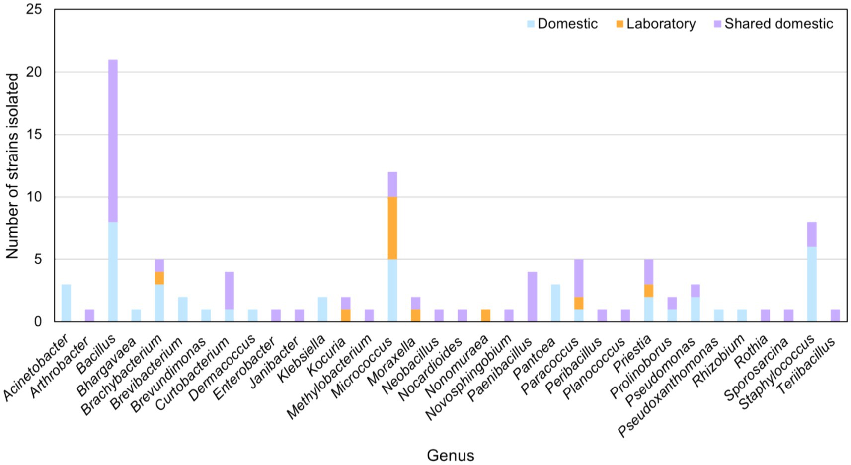
Figure 1 . Main bacterial genera isolated from domestic, domestic-shared and laboratory microwaves.
Moreover, microbial strains of the genera Bacillus , Curtobacterium , Prolinoborus , Pseudomonas , and Staphylococus were isolated from both domestic and domestic-shared microwaves. Kocuria and Moraxella strains were obtained from domestic-shared and laboratory microwaves. Members of four genera were found in all types of microwaves: Brachybacterium , Micrococcus , Paracoccus , and Priestia ( Figure 1 ).
2.2 Analysis of bacterial diversity of microwaves by NGS
NGS (Next Generation Sequencing) analysis of the conserved V3 and V4 regions of the 16S rRNA gene allowed the exploration of bacterial diversity within microwave ovens. The results showed that, at the phylum level, Proteobacteria predominated in microwave bacterial communities, followed by Firmicutes and Actinobacteria to a lesser extent ( Figure 2 ; Supplementary file S1 ). Differential abundance analysis confirmed the higher presence of the phyla Chloroflexi , Acidobacteria , Deinococcus-Thermus , and Cyanobacteria in the laboratory microwaves compared to the household microwaves ( Supplementary file S2 ). The latter phylum was also more abundant in the domestic-shared microwave group compared to the domestic (not shared) microwaves.
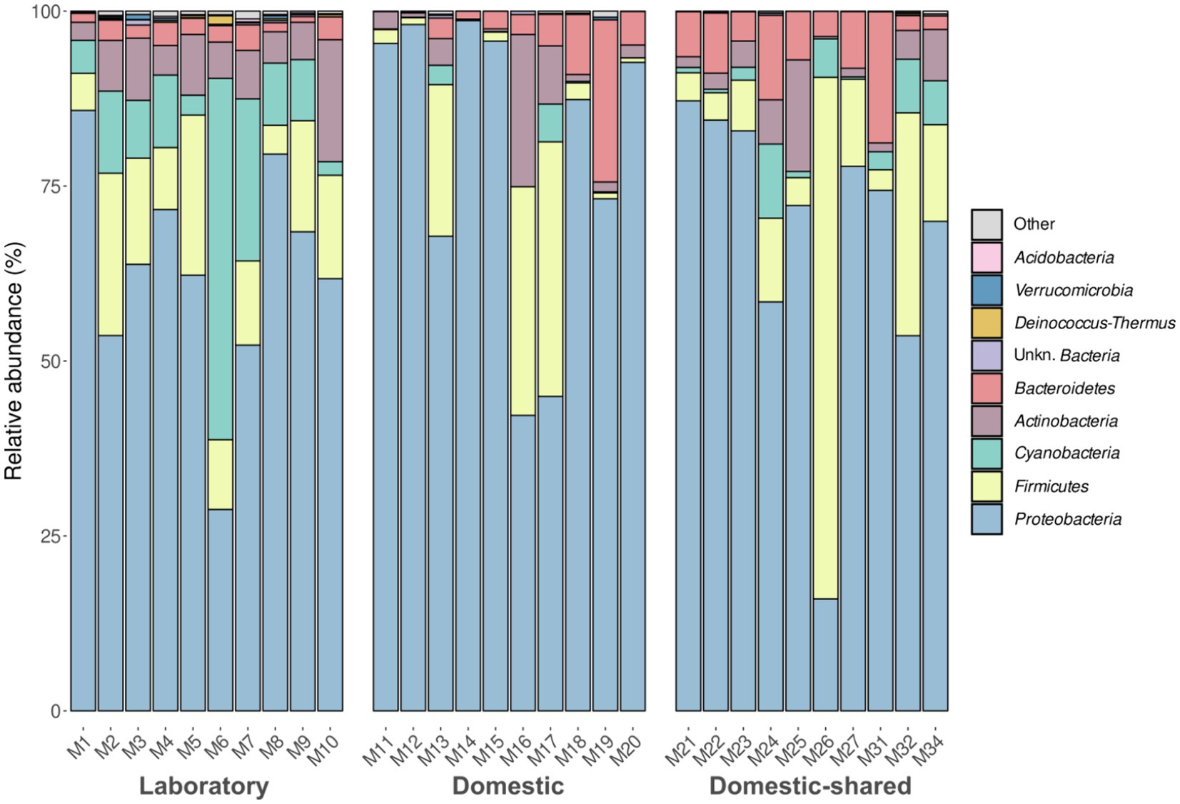
Figure 2 . Taxonomic distribution at the phylum level of the bacteria present in the three types of microwaves: laboratory (M1–M10), domestic (M11–M20), and domestic-shared (M21–M34).
At the genus level, laboratory microwaves showed a more homogeneous composition than domestic microwaves ( Figure 3 ). Acinetobacter , Pseudomonas , and Sphingobium were present in all types of microwaves. Among the significantly more abundant genera in laboratory microwaves compared to household microwaves were Delftia , Micrococcus , Deinocococcus , and an unidentified genus of the phylum Cyanobacteria ( Supplementary file S2 ). The opposite trend was observed for the genera Epilithonimonas , Klebsiella , Shewanella , and Aeromonas , among others. In addition, differential abundance analysis between domestic and domestic-shared microwaves showed that two genera, Lawsonella and Methyloversatilis , were significantly more abundant in the latter group. When comparing NGS results with the culturing techniques, it was found that almost all of the isolated genera were detected by 16S rRNA gene sequencing. Interestingly, Bhargavaea , Janibacter , and Nonomuraea , which could be cultured, were not detected by sequencing.
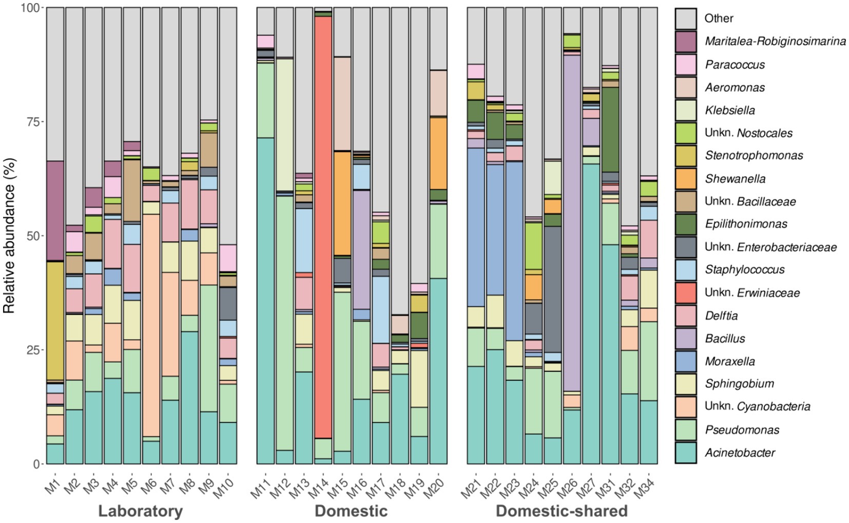
Figure 3 . Taxonomic distribution at the genus level of the bacteria present in the three types of microwaves: laboratory (M1–M10), domestic (M11–M20), and domestic-shared (M21–M34).
In terms of alpha diversity analysis, domestic microwaves had the lowest number of distinct ASVs detected and also lower Shannon index values, although these trends were only significant when comparing this type of sample with laboratory microwaves ( Figure 4 ). No significant differences were found between domestic and domestic-shared microwaves, nor between the latter and laboratory microwaves, in the number of distinct ASVs observed, Shannon index and Simpson index. Overall, between 100 and 300 different ASVs were detected, depending on the type of sample, as well as Shannon indices below 4 in household microwaves and above in laboratory microwaves, while Simpson indices ranged from 0.8 to 1.
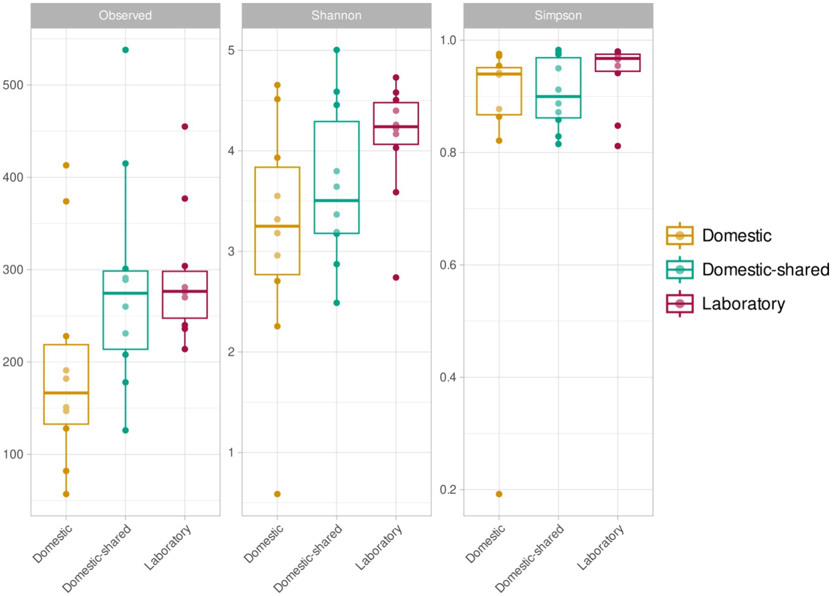
Figure 4 . Alpha diversity results (richness or number of ASVs, Shannon index and Simpson index) for the three types of microwaves: laboratory (M1–M10), domestic (M11–M20), and domestic-shared (M21–M34).
At the β-diversity level, when comparing the different groups of samples at a qualitative and quantitative level, it was observed that they were statistically different from each other (PERMANOVA test, p -value < 0.05). Laboratory samples grouped closely together, indicating a greater homogeneity in their bacterial composition ( Figure 5 ). When comparing household microwaves, samples tended to cluster within each of the two groups (domestic and shared-domestic), although this was less evident than with laboratory microwaves. Furthermore, the β-diversity of the microwave samples was also compared with that of two highly irradiated, extreme environments: solar panels and nuclear waste samples; as well as an anthropized indoor environment: kitchen surfaces ( Figure 6 ). The samples were grouped according to their origin, although the solar panel samples and especially the kitchen samples appeared to display a more similar bacterial composition to the household microwave samples. The nuclear waste disposal samples showed the least similarity to the microwave samples.
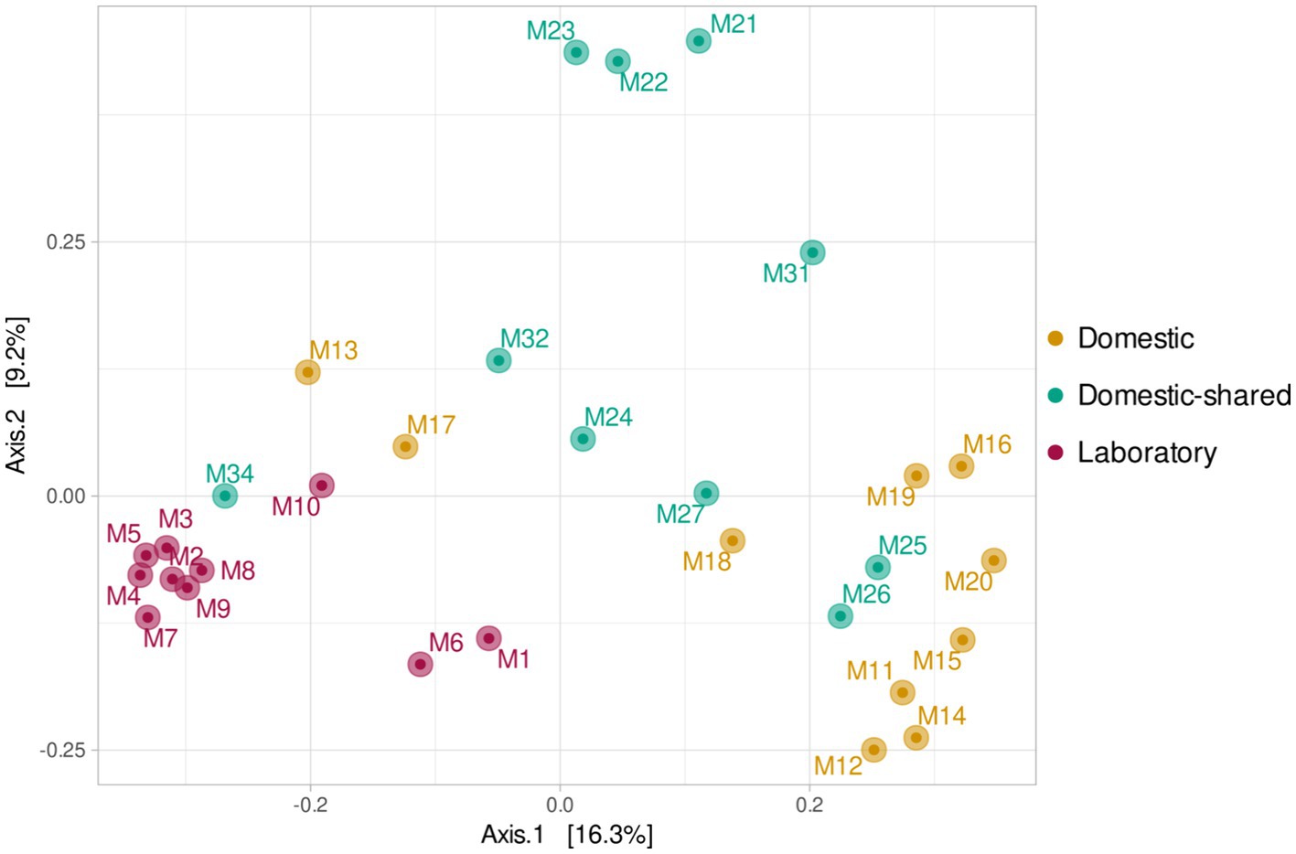
Figure 5 . Beta diversity (PCoA) based on Bray–Curtis (ASV level) for the three types of microwaves: laboratory (M1–M10), domestic (M11–M20), and domestic-shared (M21–M34).
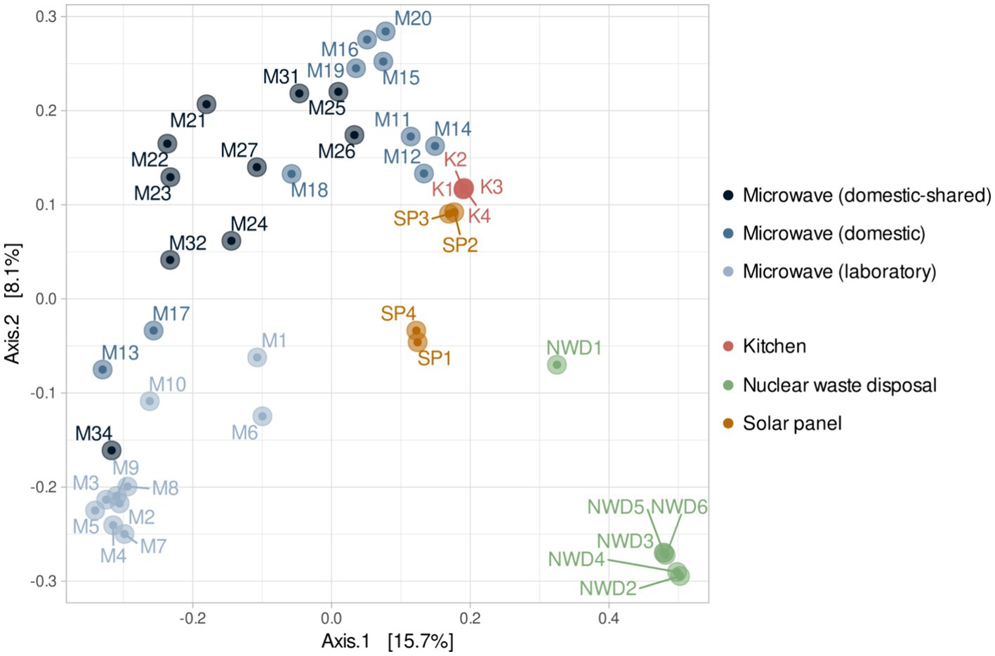
Figure 6 . Beta diversity (PCoA) based on Bray–Curtis (ASV level) for the three types of microwaves: laboratory (M1–M10), domestic (M11–M20), and domestic-shared (M21–M34) and samples from other studies: four kitchen samples, four samples from solar panels and six from nuclear waste.
3 Discussion
In this study, we describe the bacterial communities of microwaves by NGS and compare the results obtained in domestic microwaves, domestic use microwaves located in large shared spaces, and laboratory microwaves. In parallel, this work was complemented with the isolation of culturable microorganisms from the same samples.
Through culturing techniques, we found that many of the isolated strains belonged to typically commensal and anthropic genera such as Bacillus , Micrococcus , Staphylococcus , Micrococcus , and Brachybacterium ( Moskovicz et al., 2021 ; Skowron et al., 2021 ; Boxberger et al., 2022 ). As might be expected, human skin-related microorganisms are often found on artificial devices with which humans have frequent contact ( Fujiyoshi et al., 2017 ). In addition, strains belonging to genera potentially pathogenic to humans, such as Klebsiella or Brevundimonas , were identified in some samples ( Podschun and Ullmann, 1998 ; Ryan and Pembroke, 2018 ). Although these genera are less common on the skin, they can be found in the human microbiome on mucosal surfaces ( Paczosa and Mecsas, 2016 ; Leung et al., 2019 ).
Analysis of the 16S rRNA gene revealed that the bacterial communities of the microwaves were dominated by members of the phyla Proteobacteria , Firmicutes , Actinobacteria , and Bacteroidetes , which also correspond to the predominant phyla in the human skin microbiome ( Cho and Blaser, 2012 ), serving as an indicator of microwave anthropization. In this regard, the relevant presence of taxa that can be found in human skin such as Acinetobacter , Pseudomonas , Moraxella, Bacillus , and Staphylococcus ( Kumar et al., 2019 ) was also detected at the genus level. Despite the similarities found between the samples due to the frequent use of microwaves by humans, differences were also detected between the three types of microwaves, especially between laboratory and domestic microwaves. In the latter, an enrichment of food-associated genera was anticipated due to their primary culinary application. Consequently, it was logical to observe more abundant genera such as Shewanella , Enterobacter , Aeromonas , Lactococcus , or Klebsiella in this type of microwaves, as they are frequently detected in food matrices and food-related habitats, typically associated with degradation or spoilage processes ( Jarvis et al., 2018 ). It is important to note that certain species belonging to some of these genera, such as A. hydrophila , K. pneumoniae , and E. cloacae , are common contaminants in various food-related habitats and they pose potential health risks due to their pathogenic properties and antibiotic resistance ( Daskalov, 2006 ; Shaker et al., 2007 ; Rodrigues et al., 2022 ). Their presence in the microwaves, as well as on other surfaces in the built environment, suggests the importance of regular cleaning practices to mitigate potential health risks, as frequent and adequate cleaning with appropriate disinfectants helps to prevent the presence of pathogens associated with these domestic environments ( Carstens et al., 2022 ). As for laboratory microwaves, their use is completely different, as they are never used to heat food, but mainly to heat aqueous solutions, biological samples, synthetic materials or chemical reagents. Since food cannot be a shaping factor of their microbiomes, we hypothesize that the primary factor determining the microbiome in laboratory microwaves is the extreme conditions created within them (with heating processes that often require longer exposure times). In fact, some of the genera that were significantly more abundant in this group of samples included species known for their resistance to high doses of radiation, such as Deinococcus , Hymenobacter , Kineococcus , Sphingomonas , and Cellulomonas ( Nayak et al., 2021 ). Some of the mechanisms used by bacteria to withstand such adverse conditions include expression of heat shock proteins (HSPs) ( Maleki et al., 2016 ) and antioxidant enzymes ( Munteanu et al., 2015 ), maintenance of cell integrity through changes in membrane fatty acid composition ( Chen and Gänzle, 2016 ), biofilm formation ( Bogino et al., 2013 ), or DNA repair ( Sghaier et al., 2008 ). In particular, Deinococcus species such as D. radiodurans and D. geothermalis are known for their ability to withstand extreme environmental conditions such as ionizing radiation, desiccation, or high temperatures due to their highly efficient DNA repair mechanisms and protective cellular components ( Mattimore and Battista, 1996 ; Liedert et al., 2012 ). Moreover, a previous study by Shen et al. (2020) showed that Acidovorax and Aquabacterium , two other genera enriched in laboratory samples, were differentially more abundant than others at higher temperatures. The phylum Cyanobacteria and Chloroflexi , which were also more common in laboratory microwaves, have also been described as extremophiles that can withstand environments with high levels of radiation and temperature ( Lacap et al., 2011 ; Uribe-Lorío et al., 2019 ). The greater presence of bacteria resistant to these types of selective pressures could explain the higher alpha diversity values found in laboratory versus domestic microwaves. In addition, the more frequent use of domestic-shared microwaves and by more people could also favor greater diversity in this group with respect to domestic microwaves, as seen in other devices like washing machines ( Jacksch et al., 2021 ).
In addition, when the bacterial communities of microwaves were compared with those of other highly irradiated environments—solar panels and nuclear waste residues—and kitchens (food-related habitats in constant contact with humans), it was found that domestic microwaves were more similar to kitchen surface samples. However, laboratory microwaves appeared to have similarities to kitchen and, to a lesser extent, solar panel samples. Thus, genera such as Acinetobacter , Pseudomonas , Bacillus , and Staphylococcus , widely present in the vast majority of microwaves analyzed, are typical of kitchens ( Speirs et al., 1995 ; Malta et al., 2020 ). Interestingly, many of the genera significantly more present in laboratory microwaves (such as Deinococcus , Hymenobacter , Sphingomonas , Ralstonia , or Micrococcus ) are typically identified in solar panels ( Porcar et al., 2018 ; Tanner et al., 2018 , 2020 ). These results confirm that all microwave samples resembled each other, although the laboratory microwaves showed greater similarities with microbiomes from environments with relatively low organic matter and subjected to intense radiation or desiccation.
Further work is needed to study the microbial adaptations of strains isolated from microwaves to high temperatures, desiccation, and electromagnetic radiation. For example, although the ability of bacteria to tolerate high temperatures can greatly vary depending on species and strains, those present in higher abundance in microwaves - Acinetobacter , Pseudomonas , Delftia , Bacillus , and Sphingobium - are known to exhibit a range of tolerance to high temperatures, where Acinetobacter has been reported to tolerate up to 50°C ( Hrenovic et al., 2014 ), Pseudomonas up to 45°C ( Silby et al., 2009 ), Delftia up to 40°C ( Roy and Roy, 2019 ), Bacillus up to 80°C ( Thomas, 2012 ) and Sphingobium up to 40°C ( Singh et al., 2023 ). Some strains of Acinetobacter and Pseudomonas have been found to survive for extended periods of time in dry environments, including hospital surfaces ( Espinal et al., 2012 ) and air filters (Pinna et al., 2009) respectively, while Bacillus species are well-known for their ability to form spores that can survive in a desiccated state for many years ( Checinska et al., 2015 ). Similarly, some species of Sphingobium have been found to survive in dry soil and sediment environments ( Madueño et al., 2018 ).
4 Conclusion
Three types of microwaves were studied in order to shed light on their bacterial communities. Our findings revealed the intricate interplay between microwave radiation exposure, food interactions, and user habits in shaping the bacteriome of microwaves. The distinct microbial composition observed between laboratory and household microwaves underscored the influence of usage patterns on microbial communities. Household microwaves, enriched in food-associated genera, reflected their primary culinary use, while laboratory microwaves harbored radiation-, desiccation-, and high-temperature-resistant taxa, indicating prolonged exposure to microwave radiation and suggesting a selective pressure of such harsh factors in shaping the distinctive microbial profile we found. However, more research is needed to understand how certain bacterial strains commonly found in microwaves adapt to these selective pressures. Indeed, this analysis could provide relevant information regarding the biotechnological potential of the microwave bacteriome.
5 Experimental procedures
5.1 sampling.
The inner cubicle of 10 domestic, 10 shared-domestic and 10 laboratory microwaves was sampled by rubbing a sterile collection swab humidified with Phosphate Buffer Saline solution (PBS, composition in g l-1: NaCl; 8.0, KCl; 2.0, Na 2 HPO 4 ; 1.44, KH 2 PO 4 ; 0.24. pH; 7.4) that was stored in Eppendorf tubes containing 500 μL PBS and transported to the laboratory at ambient temperature (20–25°C). Samples were immediately used for strain isolation and stored at −20°C until genomic DNA was extracted. A detailed list of the samples taken, and the corresponding microwaves characteristics can be found in Supplementary Table S1 .
5.2 Strain isolation and identification
For bacterial isolation through culturing techniques, five different growth media were used in this study: Nutrient Agar (NA, composition in g/L: peptone 5, meat extract 3, NaCl 5, agar 15, pH 7.2), Reasoner’s 2A agar (R2A, composition in g/L: peptone 1, yeast extract 0.5, dextrose 0.5, soluble starch 0.5, K 2 HPO 4 0.3, MgSO 4 0.05, sodium pyruvate 0.3, 15 agar, pH 7.2), Trypticase Soy Agar medium (TSA, contained in g/L: tryptone 15, soya peptone 5, NaCl 5, agar 15, pH 7.2), Yeast Mold Agar medium (YM, contained in g/L: yeast extract 3, malt extract 3, dextrose 10, peptone soybean 4, agar 15, pH; 6.2), Columbia Blood Agar medium (CBA, contained in g/L: special peptone 23, starch 1, NaCl 5, agar 10, pH 7.3).
Samples were homogenized in Eppendorf tubes by vigorously mixing with a vortex, and serial dilutions were plated on the media above and incubated at room temperature for 7 days. After 1 week of incubation, individual colonies were selected and isolated by re-streaking onto fresh medium. Pure cultures were cryo-preserved at −80°C in 15% glycerol.
For the taxonomic identification of the strains, PCRs amplifying a fragment of the 16S rRNA gene were carried out using the universal primers 8F (5′-AGA GTT TGA TCC TGG CTC AG-3′) and 1492R (5′-CGG TTA CCT TGT TAC GAC TT-3′) after extracting the DNA by boiling the cells at 99°C for 10 min in MilliQ-water. The 16S rRNA PCR was performed using the NZYTaq II 2× Green Master Mix, and the following PCR cycle: initial denaturation at 95°C for 3 min; 30 cycles of amplification (15 s at 94°C, 15 s at 50°C, 50 s at 72°C); and 2 min of extension at 72°C. The PCR products were checked by electrophoresis in a 1.2% agarose gel and subsequently precipitated overnight in isopropanol 1:1 (vol:vol) and potassium acetate 1:10 (vol:vol; 3 M, pH 5). DNA pellets were washed with 70% ethanol, resuspended in 15 μL Milli-Q water and Sanger sequenced by Eurofins Genomics (Germany). All the sequences were manually trimmed before comparing them against the EzBioCloud 1 and NCBI online databases. 2 EzBioCloud was used to taxonomically identify the closest type strains.
5.3 Isolation of genomic DNA
Genomic DNA was isolated from the samples using the PowerSoil DNA Isolation kit (MO BIO laboratories, Carlsbad, CA, United States) following the manufacturer’s instructions and quantified using the Qubit dsDNA HS Assay kit (Qubit 2.0 Fluorometer, Q32866). Three DNA extractions of new, unused sterile collection swabs humidified with PBS solution were also carried out, one of them together with the microwave’s samples and the remaining two on different subsequent days. These two later ones were sent for high-throughput rRNA sequencing separately in two other sequencing batches with samples belonging to other projects.
5.4 High-throughput rRNA sequencing and metataxonomic analysis
In order to study the bacterial communities present in the microwaves, the extracted genomic DNA was used to amplify the hypervariable region V3-V4 of the 16S ribosomal RNA gene. The conserved regions V3 and V4 (459 bp) of the 16S rRNA gene were amplified using the following forward and reverse primers: 5′-TCG TCG GCA GCG TCA GAT GTG TAT AAG AGA CAG CCT ACG GGN GGC WGC AG 3′ and 5′-GTC TCG TGG GCT CGG AGA TGT GTA TAA GAG ACA GGA CTA CHV GGG TAT CTA ATC C-3′, and the following PCR cycle: initial denaturation at 95°C for 3 min; 25 cycles of amplification (30 s at 95°C, 30 s at 55°C, 30 s at 72°C); and 5 min of extension at 72°C ( Satari et al., 2020 ). The amplification was carried out using the KAPA HiFi HotStart ReadyMix PCR kit (KK2602). The 16S rRNA amplicons were mixed with Illumina sequencing barcoded adaptors (Nextera XT index kit v2, FC-131-2001), and libraries were normalized and merged. The pools with indexed amplicons were loaded onto the MiSeq reagent cartridge v3 (MS-102-3003) and spiked with 10% PhiX control to improve the sequencing quality, that was finally conducted using paired-ends on an Illumina MiSeq platform (2 × 300 bp) in the Foundation for the Promotion of Health and Biomedical Research of the Valencian Community (Fisabio) (Valencia, Spain).
The raw Illumina sequences were loaded into Qiime2 (v2021.2.0) ( Bolyen et al., 2019 ). The quality of the sequences was checked using the plugin Demux and the Qiime2-integrated DADA2 pipeline was used for trimming and joining the sequences, removing chimeras and detecting amplicon sequence variants (ASVs) (>99.9% of similarity). The taxonomy of each sequence variant was determined via the classify-Sklearn module from the feature-classifier plugin, employing Greengenes-SILVA-RDP (GSR) ( Molano et al., 2024 ) as reference database for the 16S rRNA taxonomic assignment (V3-V4 hypervariable region). Results were analyzed and plotted with the phyloseq R package (v. 1.30.0) ( McMurdie and Holmes, 2013 ) and ggplot2 (v3.4.0).
The beta diversity analysis was carried out using the principal component analysis (PCoA) after calculating the distances between samples using the Bray-Curtis method, using phyloseq R package (v. 1.22.3) ( McMurdie and Holmes, 2013 ) with Bray–Curtis dissimilarities. PERMANOVA tests were calculated with vegan using the adonis2 function from the vegan R package (v2.6.4) to detect statistically significant differences in the composition of the microbiome between the groups analyzed. The differential abundance analyses between taxa were conducted using the MaAsLin2 R package (v1.0.0) (Mallick et al., 2021) with the following parameters: min_abundance = 0.01, min_prevalence = 0.33, max_significance = 0.05, normalization = “None,” transform = “LOG,” analysis_method = “LM,” correction = “BH,” standardize = FALSE. Differentially abundant taxa were considered significant if the adjusted p -value was less than or equal to 0.05.
Additionally, the bacterial profile obtained in terms of β-diversity was compared with two extreme environments with high levels of radiation: solar panels and nuclear waste samples, along with a human-modified indoor environment represented by kitchen samples ( Supplementary Table S2 ). For this purpose, publicly available datasets were downloaded from NCBI.
Data availability statement
Raw reads of the samples analyzed in this study are available at NCBI’s Sequence Read Archive (SRA) (Bioproject Accession PRJNA977132).
Author contributions
AI: Conceptualization, Data curation, Formal analysis, Investigation, Methodology, Supervision, Validation, Visualization, Writing – original draft, Writing – review & editing. LM: Data curation, Formal analysis, Investigation, Methodology, Writing – review & editing. DT: Data curation, Formal analysis, Investigation, Methodology, Visualization, Writing – original draft, Writing – review & editing. MP: Conceptualization, Funding acquisition, Investigation, Project administration, Resources, Supervision, Validation, Writing – original draft, Writing – review & editing.
The author(s) declare that financial support was received for the research, authorship, and/or publication of this article. Financial support from the European Union H2020 (MIPLACE project ref. PCI2019-111845-2, Natural and Synthetic Microbial Communities for Sustainable Production of Optimised Biogas, MICRO4BIOGAS, Grant agreement ID: 101000470) and the Agencia Estatal de Investigación (AEI) (427 Programación Conjunta Internacional 2019) is acknowledged.
Conflict of interest
DT and MP were employed by Darwin Bioprospecting Excellence S.L.
The remaining authors declare that the research was conducted in the absence of any commercial or financial relationships that could be construed as a potential conflict of interest.
Publisher’s note
All claims expressed in this article are solely those of the authors and do not necessarily represent those of their affiliated organizations, or those of the publisher, the editors and the reviewers. Any product that may be evaluated in this article, or claim that may be made by its manufacturer, is not guaranteed or endorsed by the publisher.
Supplementary material
The Supplementary material for this article can be found online at: https://www.frontiersin.org/articles/10.3389/fmicb.2024.1395751/full#supplementary-material
1. ^ https://www.ezbiocloud.net
2. ^ https://blast.ncbi.nlm.nih.gov/Blast.cgi
Bogino, P., Abod, A., Nievas, F., and Giordano, W. (2013). Water-limiting conditions alter the structure and biofilm-forming ability of bacterial multispecies communities in the alfalfa rhizosphere. PLoS One 8:e79614. doi: 10.1371/journal.pone.0079614
PubMed Abstract | Crossref Full Text | Google Scholar
Bolyen, E., Rideout, J. R., Dillon, M. R., Bokulich, N. A., Abnet, C. C., Al-Ghalith, G. A., et al. (2019). Reproducible, interactive, scalable and extensible microbiome data science using QIIME 2. Nat. Biotechnol. 37, 852–857. doi: 10.1038/s41587-019-0209-9
Crossref Full Text | Google Scholar
Boxberger, M., Magnien, S., Antezack, A., Rolland, C., Makoa, M., La-Scola, B., et al. (2022). Brachybacterium epidermidis Sp. Nov., a novel bacterial species isolated from the Back of the right hand, in a 67-year-old healthy woman. Int. J. Microbiol. 2022, 2875994–2875998. doi: 10.1155/2022/2875994
Cao, J.-X., Wang, F., Li, X., Sun, Y.-Y., Wang, Y., Ou, C.-R., et al. (2018). The influence of microwave sterilization on the ultrastructure, permeability of cell membrane and expression of proteins of Bacillus Cereus . Front. Microbiol. 9:1870. doi: 10.3389/fmicb.2018.01870
Carstens, C. K., Salazar, J. K., Sharma, S. V., Chan, W., and Darkoh, C. (2022). Evaluation of the kitchen microbiome and food safety behaviors of predominantly low-income families. Front. Microbiol. 13:987925. doi: 10.3389/fmicb.2022.987925
Checinska, A., Paszczynski, A., and Burbank, M. (2015). Bacillus and other spore-forming genera: variations in responses and mechanisms for survival. Annu. Rev. Food Sci. Technol. 6, 351–369. doi: 10.1146/annurev-food-030713-092332
Chen, Y. Y., and Gänzle, M. G. (2016). Influence of cyclopropane fatty acids on heat, high pressure, acid and oxidative resistance in Escherichia coli . Int. J. Food Microbiol. 222, 16–22. doi: 10.1016/j.ijfoodmicro.2016.01.017
Cho, I., and Blaser, M. J. (2012). The human microbiome: at the interface of health and disease. Nat. Rev. Genet. 13, 260–270. doi: 10.1038/nrg3182
Daskalov, H. (2006). The importance of Aeromonas hydrophila in food safety. Food Control 17, 474–483. doi: 10.1016/j.foodcont.2005.02.009
Espinal, P., Martí, S., and Vila, J. (2012). Effect of biofilm formation on the survival of Acinetobacter baumannii on dry surfaces. J. Hosp. Infect. 80, 56–60. doi: 10.1016/j.jhin.2011.08.013
Fujiyoshi, S., Tanaka, D., and Maruyama, F. (2017). Transmission of airborne Bacteria across built environments and its measurement standards: a review. Front. Microbiol. 8:2336. doi: 10.3389/fmicb.2017.02336
Gohli, J., Bøifot, K. O., Moen, L. V., Pastuszek, P., Skogan, G., Udekwu, K. I., et al. (2019). The subway microbiome: seasonal dynamics and direct comparison of air and surface bacterial communities. Microbiome 7:160. doi: 10.1186/s40168-019-0772-9
Hrenovic, J., Durn, G., Goic-Barisic, I., and Kovacic, A. (2014). Occurrence of an environmental Acinetobacter baumannii strain similar to a clinical isolate in Paleosol from Croatia. Appl. Environ. Microbiol. 80, 2860–2866. doi: 10.1128/AEM.00312-14
Jacksch, S., Zohra, H., Weide, M., Schnell, S., and Egert, M. (2021). Cultivation-based quantification and identification of Bacteria at two hygienic key sides of domestic washing machines. Microorganisms 9:905. doi: 10.3390/microorganisms9050905
Jarvis, K. G., Daquigan, N., White, J. R., Morin, P. M., Howard, L. M., Manetas, J. E., et al. (2018). Microbiomes associated with foods from plant and animal sources. Front. Microbiol. 9:2540. doi: 10.3389/fmicb.2018.02540
Kandel, C. E., Simor, A. E., and Redelmeier, D. A. (2014). Elevator buttons as unrecognized sources of bacterial colonization in hospitals. Open Med. 8, e81–e86
PubMed Abstract | Google Scholar
Kubo, M. T., Siguemoto, É. S., Funcia, E. S., Augusto, P. E., Curet, S., Boillereaux, L., et al. (2020). Non-thermal effects of microwave and ohmic processing on microbial and enzyme inactivation: a critical review. Curr. Opin. Food Sci. 35, 36–48. doi: 10.1016/j.cofs.2020.01.004
Kumar, K. V., Pal, A., Bai, P., Kour, A., E, S., P, R., et al. (2019). Co-aggregation of bacterial flora isolated from the human skin surface. Microb. Pathog. 135:103630. doi: 10.1016/j.micpath.2019.103630
Lacap, D. C., Warren-Rhodes, K. A., McKay, C. P., and Pointing, S. B. (2011). Cyanobacteria and chloroflexi-dominated hypolithic colonization of quartz at the hyper-arid core of the Atacama Desert, Chile. Extremophiles 15, 31–38. doi: 10.1007/s00792-010-0334-3
Lax, S., Hampton-Marcell, J. T., Gibbons, S. M., Colares, G. B., Smith, D., Eisen, J. A., et al. (2015). Forensic analysis of the microbiome of phones and shoes. Microbiome 3:21. doi: 10.1186/s40168-015-0082-9
Leung, P. H. M., Subramanya, R., Mou, Q., Lee, K. T., Islam, F., Gopalan, V., et al. (2019). Characterization of mucosa-associated microbiota in matched Cancer and non-neoplastic mucosa from patients with colorectal Cancer. Front. Microbiol. 10:1317. doi: 10.3389/fmicb.2019.01317
Liedert, C., Peltola, M., Bernhardt, J., Neubauer, P., and Salkinoja-Salonen, M. (2012). Physiology of resistant Deinococcus geothermalis bacterium aerobically cultivated in low-manganese medium. J. Bacteriol. 194, 1552–1561. doi: 10.1128/JB.06429-11
Madueño, L., Coppotelli, B. M., Festa, S., Alvarez, H. M., and Morelli, I. S. (2018). Insights into the mechanisms of desiccation resistance of the Patagonian PAH-degrading strain Sphingobium sp. 22B. J. Appl. Microbiol. 124, 1532–1543. doi: 10.1111/jam.13742
Maleki, F., Khosravi, A., Nasser, A., Taghinejad, H., and Azizian, M. (2016). Bacterial heat shock protein activity. J. Clin. Diagn. Res. 10:BE01–BE03. doi: 10.7860/JCDR/2016/14568.7444
Mallick, H., Rahnavard, A., McIver, L. J., Ma, S., Zhang, Y., Nguyen, L. H., et al. (2021). Multivariable association discovery in population-scale meta-omics studies. PLOS Computational Biology , 17:e1009442. doi: 10.1371/journal.pcbi.1009442
Malta, R. C. R., Ramos, G. L. De, and Nascimento, J. S. (2020). From food to hospital: we need to talk about Acinetobacter spp. Germs , 10, 210–217. doi: 10.18683/germs.2020.1207
Mattimore, V., and Battista, J. R. (1996). Radioresistance of Deinococcus radiodurans : functions necessary to survive ionizing radiation are also necessary to survive prolonged desiccation. J. Bacteriol. 178, 633–637. doi: 10.1128/jb.178.3.633-637.1996
McMurdie, P. J., and Holmes, S. (2013). Phyloseq: an R package for reproducible interactive analysis and graphics of microbiome census data. PLoS One 8:e61217. doi: 10.1371/journal.pone.0061217
Molano, L.-A. G., Vega-Abellaneda, S., and Manichanh, C. (2024). GSR-DB: a manually curated and optimized taxonomical database for 16S rRNA amplicon analysis. mSystems 9, e00950–e00923. doi: 10.1128/msystems.00950-23
Moskovicz, V., Ben-El, R., Horev, G., and Mizrahi, B. (2021). Skin microbiota dynamics following B. subtilis formulation challenge: an in vivo study in mice. BMC Microbiol. 21:231. doi: 10.1186/s12866-021-02295-y
Munteanu, A.-C., Uivarosi, V., and Andries, A. (2015). Recent progress in understanding the molecular mechanisms of radioresistance in Deinococcus bacteria. Extremophiles 19, 707–719. doi: 10.1007/s00792-015-0759-9
Nayak, T., Sengupta, I., and Dhal, P. K. (2021). A new era of radiation resistance bacteria in bioremediation and production of bioactive compounds with therapeutic potential and other aspects: an in-perspective review. J. Environ. Radioact. 237:106696. doi: 10.1016/j.jenvrad.2021.106696
Paczosa, M. K., and Mecsas, J. (2016). Klebsiella pneumoniae : going on the offense with a strong defense. Microbiol. Mol. Biol. Rev. 80, 629–661. doi: 10.1128/mmbr.00078-15
Pinna, A., Usai, D., Sechi, L. A., Zanetti, S., Jesudasan, N. C. A., Thomas, P. A., et al. (2009). An Outbreak of Post-Cataract Surgery Endophthalmitis Caused by Pseudomonas aeruginosa. Ophthalmology , 116, 2321–2326.e4. doi: 10.1016/j.ophtha.2009.06.004
Podschun, R., and Ullmann, U. (1998). Klebsiella spp. as nosocomial pathogens: epidemiology, taxonomy, typing methods, and pathogenicity factors. Clin. Microbiol. Rev. 11, 589–603. doi: 10.1128/CMR.11.4.589
Porcar, M., Louie, K. B., Kosina, S. M., Van Goethem, M. W., Bowen, B. P., Tanner, K., et al. (2018). Microbial ecology on solar panels in Berkeley, CA, United States. Front. Microbiol. 9:3043. doi: 10.3389/fmicb.2018.03043
Raghupathi, P. K., Zupančič, J., Brejnrod, A. D., Jacquiod, S., Houf, K., Burmølle, M., et al. (2018). Microbial diversity and putative opportunistic pathogens in dishwasher biofilm communities. Appl. Environ. Microbiol. 84, e02755–e02717. doi: 10.1128/AEM.02755-17
Rodrigues, C., Hauser, K., Cahill, N., Ligowska-Marzęta, M., Centorotola, G., Cornacchia, A., et al. (2022). High prevalence of Klebsiella pneumoniae in European food products: a multicentric study comparing culture and molecular detection methods. Microbiol. Spectr. 10:e0237621. doi: 10.1128/spectrum.02376-21
Roy, S., and Roy, M. (2019). Characterization of plant growth promoting feature of a neutromesophilic, facultatively chemolithoautotrophic, Sulphur oxidizing bacterium Delftia sp. strain SR4 isolated from coal mine spoil. Int. J. Phytoremediation 21, 531–540. doi: 10.1080/15226514.2018.1537238
Ryan, M. P., and Pembroke, J. T. (2018). Brevundimonas spp: emerging global opportunistic pathogens. Virulence 9, 480–493. doi: 10.1080/21505594.2017.1419116
Satari, L., Guillén, A., Vidal-Verdú, À., and Porcar, M. (2020). The wasted chewing gum bacteriome. Sci. Rep. 10:16846. doi: 10.1038/s41598-020-73913-4
Sghaier, H., Ghedira, K., Benkahla, A., and Barkallah, I. (2008). Basal DNA repair machinery is subject to positive selection in ionizing-radiation-resistant bacteria. BMC Genomics 9:297. doi: 10.1186/1471-2164-9-297
Shaker, R., Osaili, T., Al-Omary, W., Jaradat, Z., and Al-Zuby, M. (2007). Isolation of Enterobacter sakazakii and other Enterobacter sp. from food and food production environments. Food Control 18, 1241–1245. doi: 10.1016/j.foodcont.2006.07.020
Shaw, P., Kumar, N., Mumtaz, S., Lim, J. S., Jang, J. H., Kim, D., et al. (2021). Evaluation of non-thermal effect of microwave radiation and its mode of action in bacterial cell inactivation. Sci. Rep. 11:14003. doi: 10.1038/s41598-021-93274-w
Shen, Q., Ji, F., Wei, J., Fang, D., Zhang, Q., Jiang, L., et al. (2020). The influence mechanism of temperature on solid phase denitrification based on denitrification performance, carbon balance, and microbial analysis. Sci. Total Environ. 732:139333. doi: 10.1016/j.scitotenv.2020.139333
Shu, W.-S., and Huang, L.-N. (2022). Microbial diversity in extreme environments. Nat. Rev. Microbiol. 20, 219–235. doi: 10.1038/s41579-021-00648-y
Silby, M. W., Nicoll, J. S., and Levy, S. B. (2009). Requirement of polyphosphate by Pseudomonas fluorescens Pf0-1 for competitive fitness and heat tolerance in laboratory media and sterile soil. Appl. Environ. Microbiol. 75, 3872–3881. doi: 10.1128/AEM.00017-09
Singh, A., Pandey, A. K., and Dubey, S. K. (2023). Genome sequencing and in silico analysis of isoprene degrading monooxygenase enzymes of Sphingobium sp. BHU LFT2. J. Biomol. Struct. Dyn. 41, 3821–3834. doi: 10.1080/07391102.2022.2057360
Skowron, K., Bauza-Kaszewska, J., Kraszewska, Z., Wiktorczyk-Kapischke, N., Grudlewska-Buda, K., Kwiecińska-Piróg, J., et al. (2021). Human skin microbiome: impact of intrinsic and extrinsic factors on skin microbiota. Microorganisms 9:543. doi: 10.3390/microorganisms9030543
Speirs, J. P., Anderton, A., and Anderson, J. G. (1995). A study of the microbial content of the domestic kitchen. Int. J. Environ. Health Res. 5, 109–122. doi: 10.1080/09603129509356839
Tanner, K., Martí, J. M., Belliure, J., Fernández-Méndez, M., Molina-Menor, E., Peretó, J., et al. (2018). Polar solar panels: Arctic and Antarctic microbiomes display similar taxonomic profiles. Environ. Microbiol. Rep. 10, 75–79. doi: 10.1111/1758-2229.12608
Tanner, K., Molina-Menor, E., Latorre-Pérez, A., Vidal-Verdú, À., Vilanova, C., Peretó, J., et al. (2020). Extremophilic microbial communities on photovoltaic panel surfaces: a two-year study. Microb. Biotechnol. 13, 1819–1830. doi: 10.1111/1751-7915.13620
Thomas, P. (2012). Long-term survival of Bacillus spores in alcohol and identification of 90% ethanol as relatively more spori/bactericidal. Curr. Microbiol. 64, 130–139. doi: 10.1007/s00284-011-0040-0
Uribe-Lorío, L., Brenes-Guillén, L., Hernández-Ascencio, W., Mora-Amador, R., González, G., Ramírez-Umaña, C. J., et al. (2019). The influence of temperature and pH on bacterial community composition of microbial mats in hot springs from Costa Rica. Microbiologyopen 8:e893. doi: 10.1002/mbo3.893
Vilanova, C., Iglesias, A., and Porcar, M. (2015). The coffee-machine bacteriome: biodiversity and colonisation of the wasted coffee tray leach. Sci. Rep. 5:17163. doi: 10.1038/srep17163
Woo, I. S., Rhee, I. K., and Park, H. D. (2000). Differential damage in bacterial cells by microwave radiation on the basis of cell wall structure. Appl. Environ. Microbiol. 66, 2243–2247. doi: 10.1128/AEM.66.5.2243-2247.2000
Keywords: microwave, 16S rRNA gene sequencing, taxonomic classification, radiation, desiccation, selective pressure
Citation: Iglesias A, Martínez L, Torrent D and Porcar M (2024) The microwave bacteriome: biodiversity of domestic and laboratory microwave ovens. Front. Microbiol . 15:1395751. doi: 10.3389/fmicb.2024.1395751
Received: 04 March 2024; Accepted: 19 June 2024; Published: 08 August 2024.
Reviewed by:
Copyright © 2024 Iglesias, Martínez, Torrent and Porcar. This is an open-access article distributed under the terms of the Creative Commons Attribution License (CC BY) . The use, distribution or reproduction in other forums is permitted, provided the original author(s) and the copyright owner(s) are credited and that the original publication in this journal is cited, in accordance with accepted academic practice. No use, distribution or reproduction is permitted which does not comply with these terms.
*Correspondence: Manuel Porcar, [email protected]
† ORCID: Alba Iglesias, http://orcid.org/0000-0002-6582-9747 Daniel Torrent, http://orcid.org/0000-0002-3997-0974 Manuel Porcar, http://orcid.org/0000-0002-7916-9479
Disclaimer: All claims expressed in this article are solely those of the authors and do not necessarily represent those of their affiliated organizations, or those of the publisher, the editors and the reviewers. Any product that may be evaluated in this article or claim that may be made by its manufacturer is not guaranteed or endorsed by the publisher.

IMAGES
COMMENTS
There are 4 schedules (or parts) in the Bio-Medical Waste rules 2016: Schedule 1: Categorization and Management. Schedule 2: Standards for treatment and disposal of BMW. Schedule 3: Prescribed Authority and duties. Schedule 4: Label of containers, bags and transportation of Bio-Medical waste.
Biomedical waste management involves the proper handling, disposal, and treatment of waste materials generated in healthcare facilities, research laboratories, and other medical settings. It refers to a collection of practises intended to reduce the risks connected with biomedical waste, such as infectious diseases and environmental damage.
Comprehensive management of biomedical waste relies on a. blend of ed ucation and awareness efforts. Education and. dissemination of knowledge ensure that healthcare personnel, waste handlers and ...
The indiscriminate dumping of biomedical wastes by hospitals and nursing homes was a source of pollution that caused dangers to the health and environment. In order to overcome this crisis, the Bio Medical Waste (Handling and Management) Rules, were notified in July 1998. The rules seek to introduce biomedical waste disposal practices in India.
waste in a safe and sustainable manner. Effective and efficient biomedical waste management is essential to achieving a cleaner environment where humans and health can flour ish.
The biomedical waste is the waste that is generated during the diagnosis, treatment or immunization of human beings or animals or in research activities pertaining thereto, or in the production or testing of biological components. Steps of biomedical waste management are a) segregation b) storage c) transport d) disposal.
Abstract The waste generated in various hospitals and healthcare facilities, including the waste of industries, can be grouped under biomedical waste (BMW). The constituents of this type of waste are various infectious and hazardous materials. This waste is then identified, segregated, and treated scientifically.
cal waste segregation and disposal. Biomedical waste management is a team task a. d responsibility of each personnel. In view of the latest guidelines and amendment biomedical waste management rules, 2018 issued by Government of India, we are providing this biomedical waste management manual to all stake.
The aim of this review article is to provide systematic evidence-based information along with a comprehensive study of BMW in an organized manner. The waste generated in various hospitals and healthcare facilities, including the waste of industries, can be grouped under biomedical waste (BMW). The constituents of this type of waste are various infectious and hazardous materials. This waste is ...
Bio-medical waste means "any solid and/or liquid waste produced during diagnosis, treatment or vaccination of human beings or animals. Biomedical waste creates hazard due to two principal reasons: infectivity and toxicity.
The issue of biomedical waste management has assumed great significance in recent times particularly in view of the rapid upsurge of HIV infection. Government of India has made proper handling and disposal of this category of waste a statutory requirement ...
BIOMEDICAL WASTE MANAGEMENT Meaning - Categories of biomedical wastes -Process of waste management. Disposal of biomedical waste products - Methods, Government Rules and regulations - Standards for Waste autoclaving, microwaving and deep burial. Agencies appointment for waste disposal.
Background: Biomedical Waste means any waste, which is generated during diagnosis, treatment or immunization of human beings or animals, or in research activities pertaining thereto or in the production or testing of biological, and including categories as mentioned in the Bio-medical Waste (Management and Handling) Rules, 1998.
The population chemical, health-care and/or hazardous microbiological associated to treatment, mainly human of waste[2]. blood, body handling, of dressings, and sharps like glasses, needles, blades, scalpels, lancets residuals of medicines, unhygienic. generated. diagnosis, treatment or [3]. Collection and disposal of biomedical waste has ...
Biomedical wastes are defined as waste that is generated during the diagnosis, treatment or immunization of human beings or animals, or in research activities pertaining thereto, or in the production of biological, and including 10 categories mentioned in Schedule I.
Introduction 'Bio-medical waste' means any waste generated during diagnosis, treatment or immunization of human beings or animals. Management of healthcare waste is an integral part of infection control and hygiene programs in healthcare settings.
Acceptable management of biomedical waste management begins from the initial stage of generation of waste, segregation at the source, storage at the site, disinfection, and transfer to the terminal disposal site plays a critical role in the disposal of waste. Hence adequate knowledge, attitudes and practices of the staff of the health care institutes play a very important role. 8,4,9
Health care waste is a unique category of waste by the source of generation, the quality of its composition, its hazardous nature and the need for appropriate protection during handling, treatment and disposal. Little knowledge and inappropriate technique of handling of biomedical waste can lead to serious consequences on health of the individual handling the bio-medical waste, the community ...
The objective of this study was to critically evaluate the existing management practices of biomedical waste and its possible health risks on our environment. A detailed study of major hospitals (Government and Private) of Bareilly city was carried out to assess the current situation of Bio-medical waste generation and management.
Items in eGyanKosh are protected by copyright, with all rights reserved, unless otherwise indicated.
Assignment Agreement V-52(5)_247573 Rental Vehicles - Short Term and Long Term Amended contact to add electric vehicles and a Premium SUV vehicle W- 131_215803 Window Washing - Facilities Management Amended to extend contract to 06/30/2025; with price schedule update W- 192(5)_215479 Waste Disposal: Infectious (Biomedical) Waste Disposal
The nuclear waste disposal samples showed the least similarity to the microwave samples. ... (2 × 300 bp) in the Foundation for the Promotion of Health and Biomedical Research of the Valencian Community (Fisabio) (Valencia, Spain). ... (GSR) (Molano et al., 2024) as reference database for the 16S rRNA taxonomic assignment (V3-V4 hypervariable ...